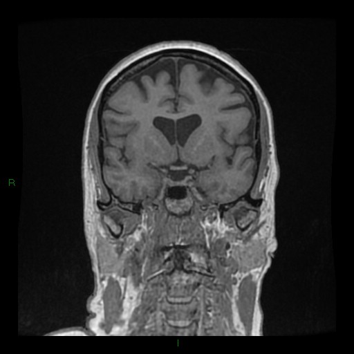
File:Cerebellar abscess (Radiopaedia 78135-90678 Coronal T1 C+ 65).jpg

From NC Commons
Jump to navigation Jump to search

Cerebellar_abscess_(Radiopaedia_78135-90678_Coronal_T1_C+_65).jpg(512 × 512 pixels, file size: 37 KB, MIME type: image/jpeg)

Summary:

Description
  • Radiopaedia case ID: 78135
  • Image ID: 52772846
  • Image stack position: 65/168
  • Plane projection: Coronal
  • Aux modality: T1 C+
  • Study findings: 4.1 x 2.8 cm rim enhancing lesion in the posteromedial aspect of the left cerebellar hemisphere. This extends towards the dural surface which is thickened and enhancing, with a tiny tract extending from the inferior wall of the abscess towards the dura and into the bony ostium.  Avid diffusion restriction within the lesion. Moderate amount of perilesional cytotoxic edema. The mass and abscess combine to displace and efface the fourth ventricle. No hydrocephalus. No other enhancing lesions. Small amount of periventricular small vessel ischemic change.
  • Modality: MRI
  • System: Central Nervous System
  • Findings: 4cm rim enhancing cystic lesion in the left cerebellar hemisphere. Moderate perilesional edema. The 4th ventricle is effaced and displaced to the right. Prominence of the temporal horns. The venous sinuses opacify normally.   Normally aerated mastoid air cells.
Date Published: 7th Jun 2020
Source https://radiopaedia.org/cases/cerebellar-abscess-5
Author Ian Bickle
Permission
(Permission-reusing-text)
http://creativecommons.org/licenses/by-nc-sa/3.0/

Licensing:

Attribution-NonCommercial-ShareAlike 3.0 Unported (CC BY-NC-SA 3.0)

File history

Click on a date/time to view the file as it appeared at that time.

Date/TimeThumbnailDimensionsUserComment
current00:43, 15 July 2021Thumbnail for version as of 00:43, 15 July 2021512 × 512 (37 KB) (talk | contribs)Radiopaedia project rID:78135 (batch #6606-245 F65)

Metadata