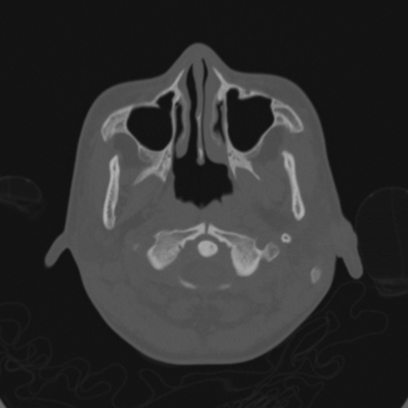
File:Cerebellar abscess secondary to mastoiditis (Radiopaedia 26284-26413 Axial bone window 6).jpg
Jump to navigation
Jump to search
Cerebellar_abscess_secondary_to_mastoiditis_(Radiopaedia_26284-26413_Axial_bone_window_6).jpg (594 × 594 pixels, file size: 52 KB, MIME type: image/jpeg)
Summary:
| Description |
|
| Date | Published: 11th Dec 2013 |
| Source | https://radiopaedia.org/cases/cerebellar-abscess-secondary-to-mastoiditis |
| Author | Ian Bickle |
| Permission (Permission-reusing-text) |
http://creativecommons.org/licenses/by-nc-sa/3.0/ |
Licensing:
Attribution-NonCommercial-ShareAlike 3.0 Unported (CC BY-NC-SA 3.0)
File history
Click on a date/time to view the file as it appeared at that time.
| Date/Time | Thumbnail | Dimensions | User | Comment | |
|---|---|---|---|---|---|
| current | 01:57, 15 July 2021 |  | 594 × 594 (52 KB) | Fæ (talk | contribs) | Radiopaedia project rID:26284 (batch #6608-6 A6) |
You cannot overwrite this file.
File usage
The following page uses this file:
