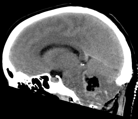
File:Cerebellar ependymoma complicated by post-operative subdural hematoma (Radiopaedia 83322-97737 C 16).png
Jump to navigation
Jump to search
Cerebellar_ependymoma_complicated_by_post-operative_subdural_hematoma_(Radiopaedia_83322-97737_C_16).png (588 × 508 pixels, file size: 131 KB, MIME type: image/png)
Summary:
| Description |
|
| Date | Published: 30th Nov 2020 |
| Source | https://radiopaedia.org/cases/cerebellar-ependymoma-complicated-by-post-operative-subdural-haematoma |
| Author | Kosuke Kato |
| Permission (Permission-reusing-text) |
http://creativecommons.org/licenses/by-nc-sa/3.0/ |
Licensing:
Attribution-NonCommercial-ShareAlike 3.0 Unported (CC BY-NC-SA 3.0)
File history
Click on a date/time to view the file as it appeared at that time.
| Date/Time | Thumbnail | Dimensions | User | Comment | |
|---|---|---|---|---|---|
| current | 08:00, 15 July 2021 |  | 588 × 508 (131 KB) | Fæ (talk | contribs) | Radiopaedia project rID:83322 (batch #6622-82 C16) |
You cannot overwrite this file.
File usage
There are no pages that use this file.
