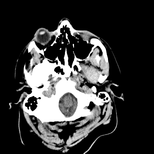
File:Cerebellar infarct due to vertebral artery dissection with posterior fossa decompression (Radiopaedia 82779-97029 Axial non-contrast 2).png
Jump to navigation
Jump to search
Cerebellar_infarct_due_to_vertebral_artery_dissection_with_posterior_fossa_decompression_(Radiopaedia_82779-97029_Axial_non-contrast_2).png (512 × 512 pixels, file size: 80 KB, MIME type: image/png)
Summary:
| Description |
|
| Date | Published: 5th Oct 2020 |
| Source | https://radiopaedia.org/cases/cerebellar-infarct-due-to-vertebral-artery-dissection-with-posterior-fossa-decompression |
| Author | Frank Gaillard |
| Permission (Permission-reusing-text) |
http://creativecommons.org/licenses/by-nc-sa/3.0/ |
Licensing:
Attribution-NonCommercial-ShareAlike 3.0 Unported (CC BY-NC-SA 3.0)
File history
Click on a date/time to view the file as it appeared at that time.
| Date/Time | Thumbnail | Dimensions | User | Comment | |
|---|---|---|---|---|---|
| current | 21:17, 15 July 2021 |  | 512 × 512 (80 KB) | Fæ (talk | contribs) | Radiopaedia project rID:82779 (batch #6651-2 A2) |
You cannot overwrite this file.
File usage
There are no pages that use this file.
