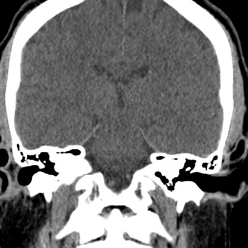
File:Cerebellar infarction (Radiopaedia 16625-16327 Coronal non-contrast 14).jpg
Jump to navigation
Jump to search
Cerebellar_infarction_(Radiopaedia_16625-16327_Coronal_non-contrast_14).jpg (512 × 512 pixels, file size: 36 KB, MIME type: image/jpeg)
Summary:
| Description |
|
| Date | Published: 4th Feb 2012 |
| Source | https://radiopaedia.org/cases/cerebellar-infarction |
| Author | David Puyó |
| Permission (Permission-reusing-text) |
http://creativecommons.org/licenses/by-nc-sa/3.0/ |
Licensing:
Attribution-NonCommercial-ShareAlike 3.0 Unported (CC BY-NC-SA 3.0)
File history
Click on a date/time to view the file as it appeared at that time.
| Date/Time | Thumbnail | Dimensions | User | Comment | |
|---|---|---|---|---|---|
| current | 23:18, 15 July 2021 |  | 512 × 512 (36 KB) | Fæ (talk | contribs) | Radiopaedia project rID:16625 (batch #6652-103 C14) |
You cannot overwrite this file.
File usage
The following page uses this file:
