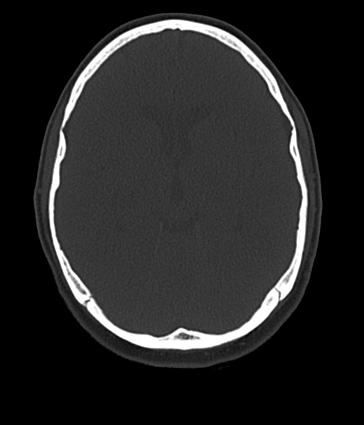
File:Cerebellar metastases - colorectal adenocarcinoma (Radiopaedia 40947-43652 AX Bone C- 2.0 MPR 29).png
Jump to navigation
Jump to search
No higher resolution available.
Cerebellar_metastases_-_colorectal_adenocarcinoma_(Radiopaedia_40947-43652_AX_Bone_C-_2.0_MPR_29).png (512 × 599 pixels, file size: 161 KB, MIME type: image/png)
Summary:
| Description |
|
| Date | Published: 18th Nov 2015 |
| Source | https://radiopaedia.org/cases/cerebellar-metastases-colorectal-adenocarcinoma |
| Author | Bruno Di Muzio |
| Permission (Permission-reusing-text) |
http://creativecommons.org/licenses/by-nc-sa/3.0/ |
Licensing:
Attribution-NonCommercial-ShareAlike 3.0 Unported (CC BY-NC-SA 3.0)
File history
Click on a date/time to view the file as it appeared at that time.
| Date/Time | Thumbnail | Dimensions | User | Comment | |
|---|---|---|---|---|---|
| current | 01:25, 16 July 2021 | | 512 × 599 (161 KB) | Fæ (talk | contribs) | Radiopaedia project rID:40947 (batch #6656-365 E29) |
You cannot overwrite this file.
File usage
The following page uses this file:
.png&oldid=767936)