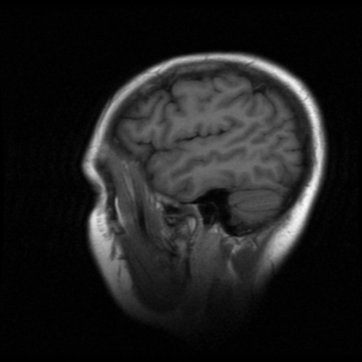
File:Cerebellar metastases - colorectal adenocarcinoma (Radiopaedia 40947-43655 Sagittal T1 4).png

From NC Commons
Jump to navigation Jump to search

Cerebellar_metastases_-_colorectal_adenocarcinoma_(Radiopaedia_40947-43655_Sagittal_T1_4).png(512 × 512 pixels, file size: 191 KB, MIME type: image/png)

Summary:

Description
  • Radiopaedia case ID: 40947
  • Image ID: 17263793
  • Image stack position: 4/20
  • Plane projection: Sagittal
  • Aux modality: T1
  • Study description: MRI Brain
  • Study findings: Technique: Multiplanar, multisequence imaging has been obtained through the brain including pre and post contrast sequences. Findings: Centered within the left inferior posterior cranial fossa, just to the left of the midline, there is a 27 mm ( craniocaudal ) x 23 mm ( AP ) x 19 mm mass which is of predominantly low signal intensity on T2-weighted imaging and isointense to grey matter on T1-weighted imaging. There is also low GRE signal. The mass demonstrates peripheral enhancement with some internal enhancing nodules mainly superiorly. The outline of the mass is slightly lobulated. There is mass effect with vasogenic edema, effacement of the cerebellar folia, mild compression of the fourth ventricle; however there is no evidence of obstructive hydrocephalus at this time. No supratentorial mass is identified. There is a small number of scattered FLAIR signal intensity within the white matter of cerebrum. Normal grey-white matter differentiation in the cerebrum. No restricted diffusion. Ventricles and basal cisterns are within normal limits. Conclusion: Solitary left posterior cranial fossa mass compatible with a hemorrhagic metastasis. Primary brain neoplasm is a possibility considered less likely.​
  • Modality: MRI
  • System: Central Nervous System
  • Findings: Technique: Pre and post contrast axial images were obtained through the brain. Findings: There is a nodular solid mass within the paramedian aspect of the left cerebellar hemisphere, spontaneously hyperdense with heterogeneous attenuation and showing vivid peripheral contrast enhancement. A mild vasogenic edema due the mass effect is noted. Despite its proximity to the fourth ventricle, there is no compression over the ventricular system. The remainder brain is unremarkable. Conclusion: A solid mass in the cerebellum most likely represents hemorrhagic metastasis, however further evaluation with MRI is advised.
Date Published: 18th Nov 2015
Source https://radiopaedia.org/cases/cerebellar-metastases-colorectal-adenocarcinoma
Author Bruno Di Muzio
Permission
(Permission-reusing-text)
http://creativecommons.org/licenses/by-nc-sa/3.0/

Licensing:

Attribution-NonCommercial-ShareAlike 3.0 Unported (CC BY-NC-SA 3.0)

File history

Click on a date/time to view the file as it appeared at that time.

Date/TimeThumbnailDimensionsUserComment
current01:32, 16 July 2021Thumbnail for version as of 01:32, 16 July 2021512 × 512 (191 KB) (talk | contribs)Radiopaedia project rID:40947 (batch #6656-4 A4)

There are no pages that use this file.
Metadata