File:Cerebellar metastasis (Radiopaedia 33616-34719 A 79).png

From NC Commons
Jump to navigation Jump to search

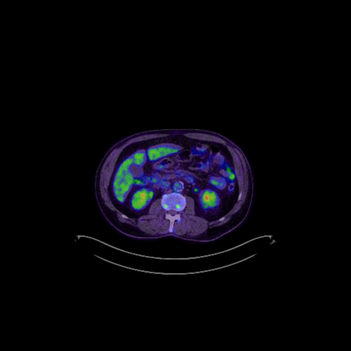
Cerebellar_metastasis_(Radiopaedia_33616-34719_A_79).png(512 × 512 pixels, file size: 110 KB, MIME type: image/png)

Summary:

Description
  • Radiopaedia case ID: 33616
  • Image ID: 10505507
  • Image stack position: 79/176
  • Plane projection: Range-AC CT WB-Tra-ALPHA Range-
  • Study findings: No evidence of lung recurrance. Right upper and middle lobectomy. Right paratracheal increased signal uptake likely nodal. Increased tracer uptake in the hepatic flexure may represent metastasis in this setting, further assessment with colonoscopy suggested.
  • Modality: Nuclear medicine
  • System: Central Nervous System
  • Findings: 35mm x 21mm x 16 mm solitary enhancing right cerebellar hemisphere lesion is identified. This contains focal internal T2 hyperintense lesions with intrinsic high T1 signal and signal loss on gradient sequences likely representing blood product (not shown). There is moderate surrounding vasogenic edema and effacement of the local sulci. Arachnoid cyst in left middle cranial fossa. Generalized cerebral volume loss. Multiple T2/FLAIR hyperintense foci are seen throughout the subcortical white matter, particularly in a periventricular location consistent with chronic small vessel ischemic change. No hydrocephalus.
Date Published: 19th Jan 2015
Source https://radiopaedia.org/cases/cerebellar-metastasis-2
Author James Sheldon
Permission
(Permission-reusing-text)
http://creativecommons.org/licenses/by-nc-sa/3.0/

Licensing:

Attribution-NonCommercial-ShareAlike 3.0 Unported (CC BY-NC-SA 3.0)

File history

Click on a date/time to view the file as it appeared at that time.

Date/TimeThumbnailDimensionsUserComment
current02:44, 16 July 2021Thumbnail for version as of 02:44, 16 July 2021512 × 512 (110 KB) (talk | contribs)Radiopaedia project rID:33616 (batch #6657-79 A79)

The following page uses this file:

Metadata