File:Cerebellar metastasis (cystic appearance) (Radiopaedia 41395-44262 Axial T1 C+ 12).png

From NC Commons
Jump to navigation Jump to search

Cerebellar_metastasis_(cystic_appearance)_(Radiopaedia_41395-44262_Axial_T1_C+_12).png(512 × 512 pixels, file size: 134 KB, MIME type: image/png)

Summary:

Description
  • Radiopaedia case ID: 41395
  • Image ID: 17786366
  • Image stack position: 12/24
  • Plane projection: Axial
  • Aux modality: T1 C+
  • Study description: MRI Brain
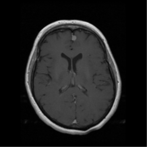
  • Study findings: Technique: Multiplanar, multisequence imaging has been obtained through the brain including pre and post contrast sequences. Findings: An irregular multicystic mass with peripheral nodular enhancement is demonstrated within the superomedial left cerebellar hemisphere. The overall lesion dimensions are 23 x 29 x 28mm (AP x trans x CC). There is moderate mass effect on the adjacent cerebellar hemisphere and vermis with FLAIR hyperintensity, as well as compression of the fourth ventricle. No hydrocephalus at this stage. No tonsillar herniation. The lesion does not demonstrate prominent flow voids. An 8mm ill-defined enhancing FLAIR hyperintense focus is demonstrated at the superolateral margin of the body of the left lateral ventricle. This does not cause significant mass effect. There is also an 8mm homogeneously enhancing extra-axial lesion abutting the anterior falx to the left of midline. Post contrast T1 volumetric acquisition through the brain for the purposes of stereotaxis has also been obtained. Conclusion: Cystic left cerebellar lesion with mass effect is most consistent with a metastasis. The enhancing focus adjacent to the left lateral ventricle is favored to also represent a metastatic deposit. The left parafalcine lesion likely represents a meningioma (metastasis less likely).
  • Modality: MRI
  • System: Central Nervous System
  • Findings: An irregular multicystic mass with peripheral nodular enhancement is demonstrated within the superomedial left cerebellar hemisphere. No hydrocephalus. A solid enhancing left frontal parafalcine nodule is also observed (meningioma?) and small nodule is noted in the left frontal corona radiata.
Date Published: 4th Dec 2015
Source https://radiopaedia.org/cases/cerebellar-metastasis-cystic-appearance
Author Bruno Di Muzio
Permission
(Permission-reusing-text)
http://creativecommons.org/licenses/by-nc-sa/3.0/

Licensing:

Attribution-NonCommercial-ShareAlike 3.0 Unported (CC BY-NC-SA 3.0)

File history

Click on a date/time to view the file as it appeared at that time.

Date/TimeThumbnailDimensionsUserComment
current09:23, 16 July 2021Thumbnail for version as of 09:23, 16 July 2021512 × 512 (134 KB) (talk | contribs)Radiopaedia project rID:41395 (batch #6665-36 B12)

There are no pages that use this file.
Metadata