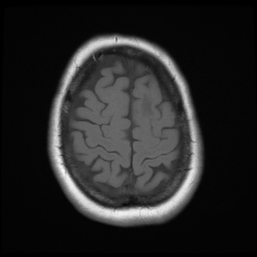
File:Cerebellar metastasis (cystic appearance) (Radiopaedia 41395-44268 Axial T1 20).png

From NC Commons
Jump to navigation Jump to search

Cerebellar_metastasis_(cystic_appearance)_(Radiopaedia_41395-44268_Axial_T1_20).png(512 × 512 pixels, file size: 171 KB, MIME type: image/png)

Summary:

Description
  • Radiopaedia case ID: 41395
  • Image ID: 17794331
  • Image stack position: 20/24
  • Plane projection: Axial
  • Aux modality: T1
  • Study description: MRI (3 months later)
  • Study findings: Since the previous exams, the 6 mm enhancing nodule in the left anterior corona radiata has developed into a multilobulated heterogeneous ring enhancing mass lesion. The right-sided midline shift of 6 mm remains stable. Mild distortion of the abutting lateral ventricle but without hydrocephalus. At the left anterior margin of the posterior fossa surgical cavity is an ovoid nodule of enhancement measuring 11 x 9 x 10 mm that is cystic inferiorly, with moderate surrounding FLAIR hyperintensity and mild mass effect on the 4th ventricle. There appear to be some tethering of this to the inferior surface of the left tentorium. More ill-defined mild-to-moderate patchy FLAIR hyperintensity surrounds the cerebellar resection cavity. Tiny focus of FLAIR hyperintensity in the right centrum semiovale, new, non-specific but most likely incidental. 8mm nodule of solid enhancement abutting the left anterior falx with a small dural tail, unchanged is more probably an incidental meningioma, although a small dural metastasis in this context remains a differential. Conclusion: Findings are in keeping with progressive metastatic disease, with a markedly enlarged left frontal mass with moderate edema. No post-operative MRI available for correlation, but a nodule of enhancement at the anterior cerebellar resection margin is in keeping with either residual and or recurrent metastasis. Unchanged since the earliest available MRI at the left anterior falx is more probably an incidental small left frontal meningioma than a dural metastasis.
  • Modality: MRI
  • System: Central Nervous System
  • Findings: An irregular multicystic mass with peripheral nodular enhancement is demonstrated within the superomedial left cerebellar hemisphere. No hydrocephalus. A solid enhancing left frontal parafalcine nodule is also observed (meningioma?) and small nodule is noted in the left frontal corona radiata.
Date Published: 4th Dec 2015
Source https://radiopaedia.org/cases/cerebellar-metastasis-cystic-appearance
Author Bruno Di Muzio
Permission
(Permission-reusing-text)
http://creativecommons.org/licenses/by-nc-sa/3.0/

Licensing:

Attribution-NonCommercial-ShareAlike 3.0 Unported (CC BY-NC-SA 3.0)

File history

Click on a date/time to view the file as it appeared at that time.

Date/TimeThumbnailDimensionsUserComment
current09:57, 16 July 2021Thumbnail for version as of 09:57, 16 July 2021512 × 512 (171 KB) (talk | contribs)Radiopaedia project rID:41395 (batch #6665-20 A20)

There are no pages that use this file.
Metadata