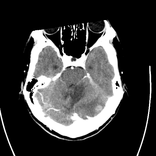
File:Cerebellar metastasis - adenocarcinoma lung (Radiopaedia 63184-71716 Axial C+ delayed 10).png
Jump to navigation
Jump to search
No higher resolution available.
Cerebellar_metastasis_-_adenocarcinoma_lung_(Radiopaedia_63184-71716_Axial_C+_delayed_10).png (512 × 512 pixels, file size: 78 KB, MIME type: image/png)
Summary:
| Description |
|
| Date | Published: 23rd Sep 2018 |
| Source | https://radiopaedia.org/cases/cerebellar-metastasis-adenocarcinoma-lung |
| Author | Frank Gaillard |
| Permission (Permission-reusing-text) |
http://creativecommons.org/licenses/by-nc-sa/3.0/ |
Licensing:
Attribution-NonCommercial-ShareAlike 3.0 Unported (CC BY-NC-SA 3.0)
File history
Click on a date/time to view the file as it appeared at that time.
| Date/Time | Thumbnail | Dimensions | User | Comment | |
|---|---|---|---|---|---|
| current | 06:16, 16 July 2021 | | 512 × 512 (78 KB) | Fæ (talk | contribs) | Radiopaedia project rID:63184 (batch #6663-41 B10) |
You cannot overwrite this file.
File usage
There are no pages that use this file.
.png&oldid=769770)