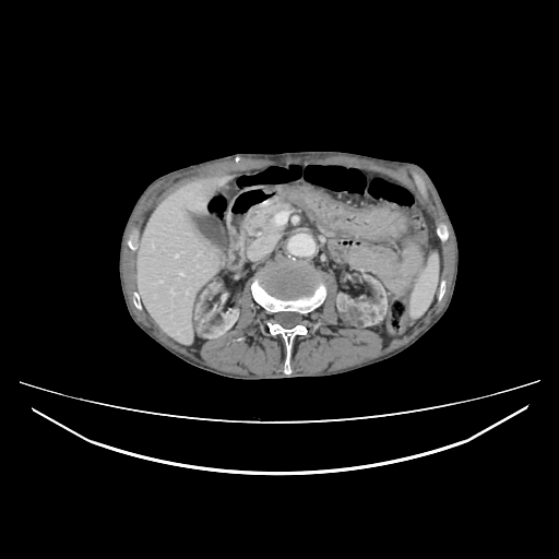
File:Cerebellar metastasis - lung tumor (Radiopaedia 69317-79150 A 42).jpg
Jump to navigation
Jump to search
Cerebellar_metastasis_-_lung_tumor_(Radiopaedia_69317-79150_A_42).jpg (512 × 512 pixels, file size: 59 KB, MIME type: image/jpeg)
Summary:
| Description |
|
| Date | Published: 12th Jul 2019 |
| Source | https://radiopaedia.org/cases/cerebellar-metastasis-lung-tumour |
| Author | Dr Ammar Haouimi |
| Permission (Permission-reusing-text) |
http://creativecommons.org/licenses/by-nc-sa/3.0/ |
Licensing:
Attribution-NonCommercial-ShareAlike 3.0 Unported (CC BY-NC-SA 3.0)
File history
Click on a date/time to view the file as it appeared at that time.
| Date/Time | Thumbnail | Dimensions | User | Comment | |
|---|---|---|---|---|---|
| current | 13:12, 16 July 2021 |  | 512 × 512 (59 KB) | Fæ (talk | contribs) | Radiopaedia project rID:69317 (batch #6667-42 A42) |
You cannot overwrite this file.
File usage
The following page uses this file:
