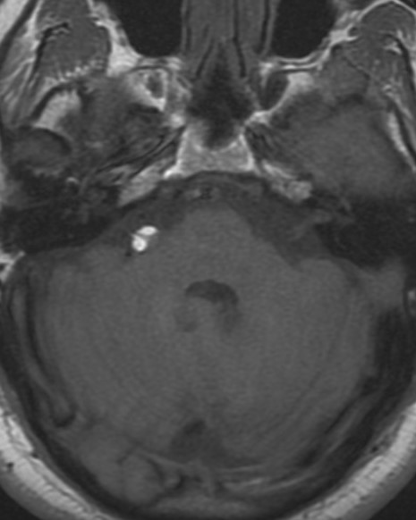
File:Cerebellopontine angle lipoma (Radiopaedia 2596-6292 Axial T1 1).jpg
Jump to navigation
Jump to search
Cerebellopontine_angle_lipoma_(Radiopaedia_2596-6292_Axial_T1_1).jpg (465 × 582 pixels, file size: 35 KB, MIME type: image/jpeg)
Summary:
| Description |
|
| Date | Published: 7th May 2008 |
| Source | https://radiopaedia.org/cases/cerebellopontine-angle-lipoma-2 |
| Author | Frank Gaillard |
| Permission (Permission-reusing-text) |
http://creativecommons.org/licenses/by-nc-sa/3.0/ |
Licensing:
Attribution-NonCommercial-ShareAlike 3.0 Unported (CC BY-NC-SA 3.0)
File history
Click on a date/time to view the file as it appeared at that time.
| Date/Time | Thumbnail | Dimensions | User | Comment | |
|---|---|---|---|---|---|
| current | 00:54, 17 July 2021 |  | 465 × 582 (35 KB) | Fæ (talk | contribs) | Radiopaedia project rID:2596 (batch #6695-1 A1) |
You cannot overwrite this file.
File usage
There are no pages that use this file.
