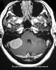
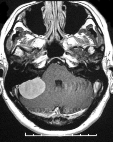
File:Cerebellopontine angle meningioma (Radiopaedia 2597-6293 Axial T1 C+ 1).jpg
Jump to navigation
Jump to search

Size of this preview: 479 × 599 pixels. Other resolutions: 192 × 240 pixels | 384 × 480 pixels.
Original file (800 × 1,001 pixels, file size: 159 KB, MIME type: image/jpeg)
Summary:
| Description |
|
| Date | Published: 7th May 2008 |
| Source | https://radiopaedia.org/cases/cerebellopontine-angle-meningioma-5 |
| Author | Frank Gaillard |
| Permission (Permission-reusing-text) |
http://creativecommons.org/licenses/by-nc-sa/3.0/ |
Licensing:
Attribution-NonCommercial-ShareAlike 3.0 Unported (CC BY-NC-SA 3.0)
File history
Click on a date/time to view the file as it appeared at that time.
| Date/Time | Thumbnail | Dimensions | User | Comment | |
|---|---|---|---|---|---|
| current | 02:01, 17 July 2021 | | 800 × 1,001 (159 KB) | Fæ (talk | contribs) | Radiopaedia project rID:2597 (batch #6706-2 B1) |
You cannot overwrite this file.
File usage
There are no pages that use this file.
.jpg&oldid=777250)