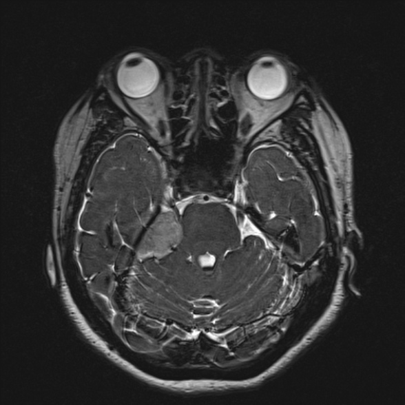
File:Cerebellopontine angle meningioma (Radiopaedia 53561-59591 Axial 3D volume T2 74).jpg
Jump to navigation
Jump to search
No higher resolution available.
Cerebellopontine_angle_meningioma_(Radiopaedia_53561-59591_Axial_3D_volume_T2_74).jpg (588 × 588 pixels, file size: 97 KB, MIME type: image/jpeg)
Summary:
| Description |
|
| Date | Published: 3rd Jun 2017 |
| Source | https://radiopaedia.org/cases/cerebellopontine-angle-meningioma-12 |
| Author | Ian Bickle |
| Permission (Permission-reusing-text) |
http://creativecommons.org/licenses/by-nc-sa/3.0/ |
Licensing:
Attribution-NonCommercial-ShareAlike 3.0 Unported (CC BY-NC-SA 3.0)
File history
Click on a date/time to view the file as it appeared at that time.
| Date/Time | Thumbnail | Dimensions | User | Comment | |
|---|---|---|---|---|---|
| current | 07:56, 17 July 2021 | | 588 × 588 (97 KB) | Fæ (talk | contribs) | Radiopaedia project rID:53561 (batch #6716-234 H74) |
You cannot overwrite this file.
File usage
The following page uses this file:
.jpg&oldid=779490)