File:Cerebral abscess with ventriculitis (Radiopaedia 78965-91878 Axial T1 23).jpg

From NC Commons
Jump to navigation Jump to search

Cerebral_abscess_with_ventriculitis_(Radiopaedia_78965-91878_Axial_T1_23).jpg(548 × 548 pixels, file size: 80 KB, MIME type: image/jpeg)

Summary:

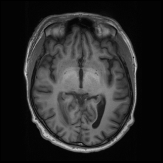
Description
  • Radiopaedia case ID: 78965
  • Image ID: 52933041
  • Image stack position: 23/50
  • Plane projection: Axial
  • Aux modality: T1
  • Study findings: Ovoid right occipital lobe white matter lesion measures 14 mm in maximum diameter. The center of the lesion is low T1 signal, reasonably high T2 signal and intermediate signal on FLAIR. There is a slightly irregular rim with immediate T1 signal and low T2 signal. Surrounding vasogenic edema extends to the occipital horn of the right lateral ventricle, where there is dependent debris filling the occipital horn with resultant loss of normal CSF signal intensity. Contrast enhancement at the periphery of the lesion anteriorly becomes contiguous with the thickened and enhancing ependymal lining of the right occipital horn and trigone. Diffusion imaging confirms true restricted diffusion in the center of the lesion as well as the dependent material in the occipital horn. No ventricular dilatation. No significant mass effect or midline shift.No other focus of signal abnormality is demonstrated. No other pathological contrast enhancement appreciated. Normal flow voids are demonstrated. The dural venous sinuses demonstrate normal contrast opacification. IMPRESSION Features in keeping with a right occipital lobe abscess and acute ventriculitis. Neurosurgical team aware of the results.
  • Modality: MRI
  • System: Central Nervous System
  • Findings: Motion artefact degrades image quality. No acute intra or extra-axial hemorrhage. There is focal hypoattenuation within the right occipital lobe, with asymmetry of the adjacent occipital horn of the right lateral ventricle and mild positive mass effect. Neuroparenchymal volume and grey-white matter differentiation are elsewhere preserved. Normal CT appearance of the midline and posterior fossa structures. Unremarkable orbits and globes. Small right frontal scalp hematoma. The imaged paranasal sinuses and mastoid air cells are well pneumatized. IMPRESSION
Date Published: 17th Jun 2020
Source https://radiopaedia.org/cases/cerebral-abscess-with-ventriculitis-2
Author Craig Hacking
Permission
(Permission-reusing-text)
http://creativecommons.org/licenses/by-nc-sa/3.0/

Licensing:

Attribution-NonCommercial-ShareAlike 3.0 Unported (CC BY-NC-SA 3.0)

File history

Click on a date/time to view the file as it appeared at that time.

Date/TimeThumbnailDimensionsUserComment
current05:41, 18 July 2021Thumbnail for version as of 05:41, 18 July 2021548 × 548 (80 KB) (talk | contribs)Radiopaedia project rID:78965 (batch #6760-23 A23)

There are no pages that use this file.