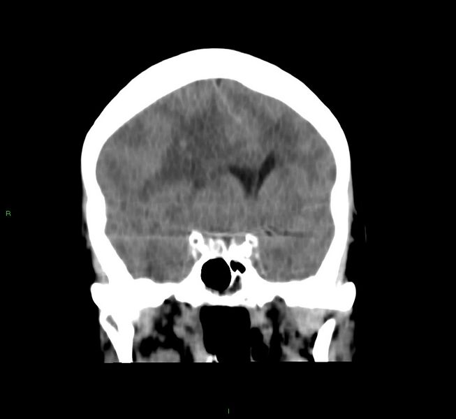
File:Cerebral amyloid angiopathy-associated lobar intracerebral hemorrhage (Radiopaedia 58751-65972 Coronal non-contrast 39).jpg
Jump to navigation
Jump to search

Size of this preview: 653 × 600 pixels. Other resolutions: 261 × 240 pixels | 522 × 480 pixels | 936 × 860 pixels.
Original file (936 × 860 pixels, file size: 55 KB, MIME type: image/jpeg)
Summary:
| Description |
|
| Date | Published: 5th Mar 2018 |
| Source | https://radiopaedia.org/cases/cerebral-amyloid-angiopathy-associated-lobar-intracerebral-haemorrhage-14 |
| Author | Mark Rodrigues |
| Permission (Permission-reusing-text) |
http://creativecommons.org/licenses/by-nc-sa/3.0/ |
Licensing:
Attribution-NonCommercial-ShareAlike 3.0 Unported (CC BY-NC-SA 3.0)
File history
Click on a date/time to view the file as it appeared at that time.
| Date/Time | Thumbnail | Dimensions | User | Comment | |
|---|---|---|---|---|---|
| current | 14:16, 21 July 2021 | | 936 × 860 (55 KB) | Fæ (talk | contribs) | Radiopaedia project rID:58751 (batch #6810-89 B39) |
You cannot overwrite this file.
File usage
The following page uses this file:
.jpg&oldid=793599)