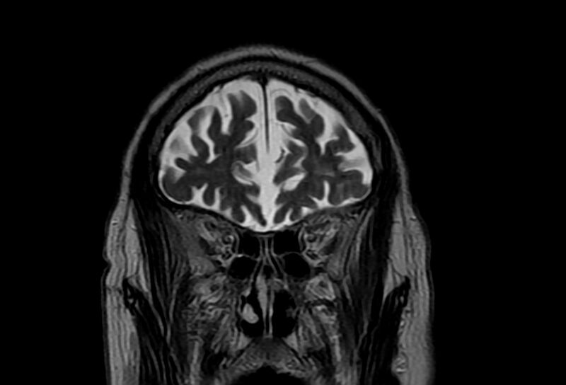
File:Cerebral amyloid angiopathy (Radiopaedia 86671-102792 Coronal T2 27).jpg
Jump to navigation
Jump to search

Size of this preview: 800 × 544 pixels. Other resolutions: 320 × 217 pixels | 640 × 435 pixels | 1,024 × 696 pixels | 1,280 × 870 pixels | 2,034 × 1,382 pixels.
Original file (2,034 × 1,382 pixels, file size: 172 KB, MIME type: image/jpeg)
Summary:
| Description |
|
| Date | Published: 8th Feb 2021 |
| Source | https://radiopaedia.org/cases/cerebral-amyloid-angiopathy-18 |
| Author | Khalid Alhusseiny |
| Permission (Permission-reusing-text) |
http://creativecommons.org/licenses/by-nc-sa/3.0/ |
Licensing:
Attribution-NonCommercial-ShareAlike 3.0 Unported (CC BY-NC-SA 3.0)
File history
Click on a date/time to view the file as it appeared at that time.
| Date/Time | Thumbnail | Dimensions | User | Comment | |
|---|---|---|---|---|---|
| current | 07:45, 18 July 2021 | | 2,034 × 1,382 (172 KB) | Fæ (talk | contribs) | Radiopaedia project rID:86671 (batch #6766-54 B27) |
You cannot overwrite this file.
File usage
There are no pages that use this file.
.jpg&oldid=788434)