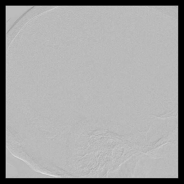
File:Cerebral aneurysm with rupture (Radiopaedia 29933-30458 LT ICA IC 28).jpg

From NC Commons
Jump to navigation Jump to search

Original file(1,024 × 1,024 pixels, file size: 126 KB, MIME type: image/jpeg)

Summary:

Description
  • Radiopaedia case ID: 29933
  • Image ID: 7485199
  • Image stack position: 28/52
  • Plane projection: LT ICA IC
  • Study findings: After informed consent, with aseptic technique, via right CFA uphill, bilateral CCA and vertebral angiograms performed, demonstrating: Large left carotid terminus aneurysm measuring 14 x 6 mm in perpendicular dimensions. During the procedure patient developed severe left sided headache with moderate confusion, with suspicion for acute rupture ( treating team was notified). Angiographic extravasation was identified. Groin was closed with a 6Fr angioseal and patient was transferred for an urgent CT. Conclusion: On table rupture of aneurysm. Large left carotid terminus saccular aneurysm as described. The aortic arch anatomy was of bovine with significant carotid tortuosity making stable position of the catheter impossible even for a SIM catheter, therefore due to likely endovascular access difficulty, the aneurysm was considered unsuitable for coil embolization.
  • Modality: DSA (angiography)
  • System: Central Nervous System
  • Findings: Brain (non-contrast): Sliver of high attenuation material within the dependent portion of the left circular sulcus likely represents a small amount of subarachnoid blood. No other intracranial hemorrhage identified. Hyperdensity seen within the suprasellar cistern corresponds to the large left terminal MCA aneurysm described below. Bilateral periventricular and deep white matter hypoattenuation is likely in keeping with chronic small vessel ischemia. Large calcified pineal gland. Ventricles and sulcal pattern are age appropriate. CTA COW: The left common carotid artery arises from the brachiocephalic trunk. The left vertebral artery arises directly from the aortic arch, between the origins of the brachiocephalic trunk and left subclavian artery. Right dominant vertebral artery. Tortuous bilateral common carotid arteries near their origins. There is a large, lobulated, anteriorly directed 11 x 4 x 14 mm aneurysm arising from the terminal left ICA, with a 3 mm neck. Further 2 mm aneurysm at the right M1 bifurcation. Left PCOM infundibulum.
Date Published: 7th Jul 2014
Source https://radiopaedia.org/cases/cerebral-aneurysm-with-rupture
Author Frank Gaillard
Permission
(Permission-reusing-text)
http://creativecommons.org/licenses/by-nc-sa/3.0/

Licensing:

Attribution-NonCommercial-ShareAlike 3.0 Unported (CC BY-NC-SA 3.0)

File history

Click on a date/time to view the file as it appeared at that time.

Date/TimeThumbnailDimensionsUserComment
current22:06, 22 July 2021Thumbnail for version as of 22:06, 22 July 20211,024 × 1,024 (126 KB) (talk | contribs)Radiopaedia project rID:29933 (batch #6835-80 B28)