File:Cerebral arterial supply to the brain - Gray's anatomy illustration (Radiopaedia 36293).jpg
Jump to navigation
Jump to search
Size of this preview: 528 × 599 pixels. Other resolutions: 211 × 240 pixels | 600 × 681 pixels.
Original file (600 × 681 pixels, file size: 158 KB, MIME type: image/jpeg)
Summary:
- Radiopaedia case ID: 36293
- Image ID: 23725
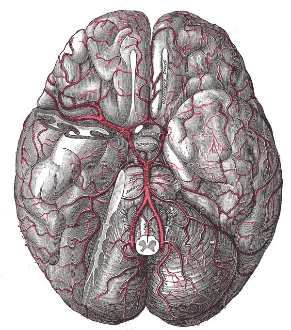
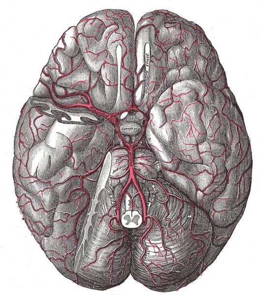
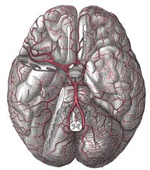
- Study findings: Cerebral arterial supply to the brain (captioned) The brain and arteries at the base of the brain. Circle of Willis is formed near the center. The temporal pole of the cerebrum and a portion of the cerebellar hemisphere have been removed on the right side. Inferior aspect (viewed from below). Diagrammatic images from Grays (20th ed): out of copyright.
- Modality: Diagram
- System: Central Nervous System
- Findings: Cerebral arterial supply to the brain (captioned)The brain and arteries at the base of the brain. Circle of Willis is formed near the center. The temporal pole of the cerebrum and a portion of the cerebellar hemisphere have been removed on the right side. Inferior aspect (viewed from below). Diagrammatic images from Grays (20th ed): out of copyright.
- Published: 16th May 2015
- Source: https://radiopaedia.org/cases/cerebral-arterial-supply-to-the-brain-grays-anatomy-illustration
- Author: Gray's Illustrations
- Permission: http://creativecommons.org/licenses/by-nc-sa/3.0/
Licensing:
Attribution-NonCommercial-ShareAlike 3.0 Unported (CC BY-NC-SA 3.0)
File history
Click on a date/time to view the file as it appeared at that time.
| Date/Time | Thumbnail | Dimensions | User | Comment | |
|---|---|---|---|---|---|
| current | 13:28, 20 March 2021 | | 600 × 681 (158 KB) | Fæ (talk | contribs) | Radiopaedia project rID:36293 (batch #6575) |
You cannot overwrite this file.
File usage
The following page uses this file:
.jpg&oldid=8859257)