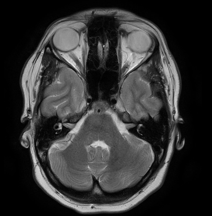
File:Cerebral arteriovenous malformation (Radiopaedia 74411-85654 Axial T2 6).jpg

From NC Commons
Jump to navigation Jump to search

Cerebral_arteriovenous_malformation_(Radiopaedia_74411-85654_Axial_T2_6).jpg(437 × 446 pixels, file size: 57 KB, MIME type: image/jpeg)

Summary:

Description
  • Radiopaedia case ID: 74411
  • Image ID: 52109736
  • Image stack position: 6/30
  • Plane projection: Axial
  • Aux modality: T2
  • Study findings: Findings: Small nidus (≤3cm) of serpiginous malformed vessels in the left posterior temporal lobe. Arterial supply is mainly from the peripheral branches (M4) of the middle cerebral artery (MCA) and less from the anterior and posterolateral choroidal arteries. Venous drainage mainly via two very prominent and multiple other smaller vessels into the superficial middle cerebral vein (SMCV), the superior anastomotic vein (vein of Trolard) and inferior anastomotic vein (vein of Labbé). Possible minor drainage via some tiny vessels into the deep veins (choroid veins). Impression: Small cerebral arteriovenous malformation in the left posterior temporal lobe with a compact nidus. Venous drainage mainly into the superficial veins. Spetzler-Martin grade 2
  • Modality: MRI
  • System: Central Nervous System
  • Findings: Serpiginous malformed vessels in the posterior temporal lobe suspicious for a cerebral arteriovenous malformation (CAVM). No other abnormalities.
Date Published: 9th Jun 2020
Source https://radiopaedia.org/cases/cerebral-arteriovenous-malformation-46
Author Joachim Feger
Permission
(Permission-reusing-text)
http://creativecommons.org/licenses/by-nc-sa/3.0/

Licensing:

Attribution-NonCommercial-ShareAlike 3.0 Unported (CC BY-NC-SA 3.0)

File history

Click on a date/time to view the file as it appeared at that time.

Date/TimeThumbnailDimensionsUserComment
current01:49, 23 July 2021Thumbnail for version as of 01:49, 23 July 2021437 × 446 (57 KB) (talk | contribs)Radiopaedia project rID:74411 (batch #6846-6 A6)

There are no pages that use this file.