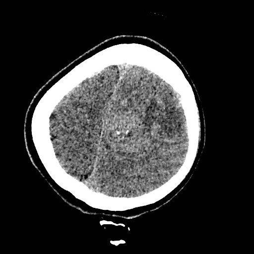
File:Cerebral arteriovenous malformation (Radiopaedia 79677-92887 Axial non-contrast 46).jpg
Jump to navigation
Jump to search
Cerebral_arteriovenous_malformation_(Radiopaedia_79677-92887_Axial_non-contrast_46).jpg (512 × 512 pixels, file size: 39 KB, MIME type: image/jpeg)
Summary:
| Description |
|
| Date | Published: 2nd Jul 2020 |
| Source | https://radiopaedia.org/cases/cerebral-arteriovenous-malformation-48 |
| Author | Mohammad Al-Tibi |
| Permission (Permission-reusing-text) |
http://creativecommons.org/licenses/by-nc-sa/3.0/ |
Licensing:
Attribution-NonCommercial-ShareAlike 3.0 Unported (CC BY-NC-SA 3.0)
File history
Click on a date/time to view the file as it appeared at that time.
| Date/Time | Thumbnail | Dimensions | User | Comment | |
|---|---|---|---|---|---|
| current | 04:45, 23 July 2021 |  | 512 × 512 (39 KB) | Fæ (talk | contribs) | Radiopaedia project rID:79677 (batch #6848-46 A46) |
You cannot overwrite this file.
File usage
The following page uses this file:
