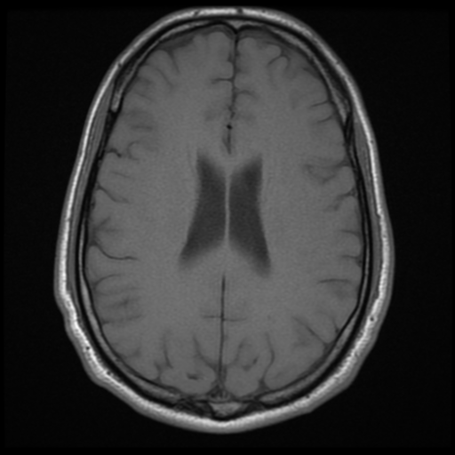
File:Cerebral arteriovenous malformation (Spetzler-Martin grade 2) (Radiopaedia 41262-44092 Axial T1 13).png

From NC Commons
Jump to navigation Jump to search

Summary:

Description
  • Radiopaedia case ID: 41262
  • Image ID: 17618986
  • Image stack position: 13/20
  • Plane projection: Axial
  • Aux modality: T1
  • Study description: MRI Brain + MRA
  • Study findings: Technique: Multiplanar and multisequence images have been obtained through the brain, including MRA and volumetric axial T1 for stereotaxic purposes. Comparison: Comparison is made with the previous CT scan. Findings: The arteriovenous malformation is again demonstrated in the posterior right superior frontal gyrus, with a compact 1.9 cm nidus in the precentral sulcus, in contact with the anterior surface of the right precentral gyrus (eloquent brain area - close to the motor cortex). The signal loss with blooming and the minimal surrounding high T2 signal are stable in appearance. Its arterial supply was previous demonstrated to be from distal branches from the right anterior and middle cerebral arteries. The venous drainage is made by cortical veins to the superior sagittal sinus, a venous varix appears present superficially. The brain parenchyma is otherwise unremarkable. No abnormal diffusion restriction or acute/subacute hematomas. The remainder circle of Willis and vertebrobasilar system opacity normally. No evidence of significant arterial stenosis or aneurysm. Conclusion: Stable appearance of the right frontal arteriovenous malformation (Spetzler-Martin grade 2).
  • Modality: MRI
  • System: Central Nervous System
  • Findings: Technique: Volumetric imaging acquisition through the brain has been obtained pre and post-contrast administration, including CT arterial angiogram. Findings: There is a right superior frontal gyrus arteriovenous malformation characterized by a compact nidus measuring 1,9 cm and being supplied by distal branches from the right ACA and right MCA, and drained by superficial cortical veins that finish at the superior sagittal sinus. There is no signs of acute hemorrhage. The remainder brain parenchyma is unremarkable. Conclusion: Right frontal arteriovenous malformation (Spetzler-Martin grade 2).
Date Published: 24th Nov 2015
Source https://radiopaedia.org/cases/cerebral-arteriovenous-malformation-spetzler-martin-grade-2
Author Bruno Di Muzio
Permission
(Permission-reusing-text)
http://creativecommons.org/licenses/by-nc-sa/3.0/

Licensing:

Attribution-NonCommercial-ShareAlike 3.0 Unported (CC BY-NC-SA 3.0)

File history

Click on a date/time to view the file as it appeared at that time.

Date/TimeThumbnailDimensionsUserComment
current00:59, 25 July 2021Thumbnail for version as of 00:59, 25 July 2021512 × 512 (200 KB) (talk | contribs)Radiopaedia project rID:41262 (batch #6890-13 A13)

There are no pages that use this file.
Metadata