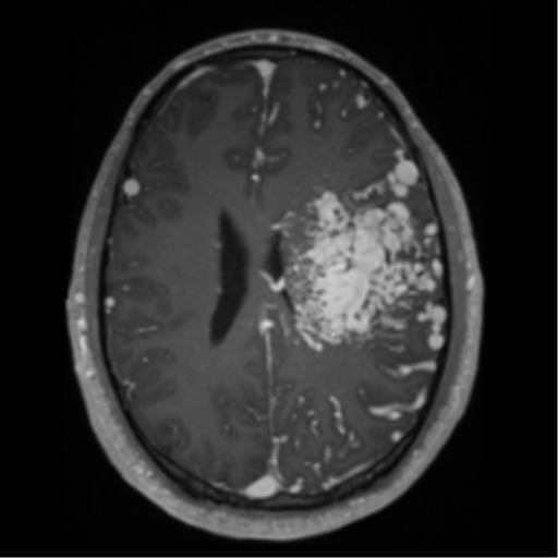
File:Cerebral arteriovenous malformation - huge (Radiopaedia 35734-37272 Axial T1 C+ 46).png
Jump to navigation
Jump to search
Cerebral_arteriovenous_malformation_-_huge_(Radiopaedia_35734-37272_Axial_T1_C+_46).png (512 × 512 pixels, file size: 165 KB, MIME type: image/png)
Summary:
| Description |
|
| Date | Published: 7th Mar 2016 |
| Source | https://radiopaedia.org/cases/cerebral-arteriovenous-malformation-huge |
| Author | Frank Gaillard |
| Permission (Permission-reusing-text) |
http://creativecommons.org/licenses/by-nc-sa/3.0/ |
Licensing:
Attribution-NonCommercial-ShareAlike 3.0 Unported (CC BY-NC-SA 3.0)
File history
Click on a date/time to view the file as it appeared at that time.
| Date/Time | Thumbnail | Dimensions | User | Comment | |
|---|---|---|---|---|---|
| current | 22:27, 24 July 2021 |  | 512 × 512 (165 KB) | Fæ (talk | contribs) | Radiopaedia project rID:35734 (batch #6887-46 A46) |
You cannot overwrite this file.
File usage
The following page uses this file:
