File:Cerebral arteriovenous malformation with lobar hemorrhage (Radiopaedia 44725-48512 Axial non-contrast 9).png

From NC Commons
Jump to navigation Jump to search

Summary:

Description
  • Radiopaedia case ID: 44725
  • Image ID: 21863760
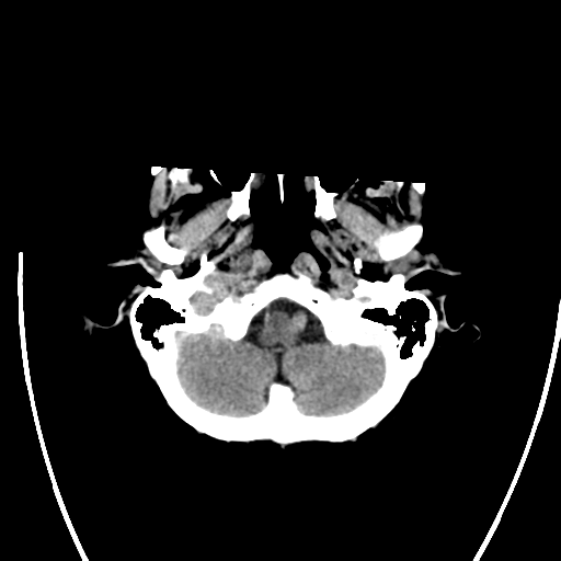
  • Image stack position: 9/34
  • Plane projection: Axial
  • Aux modality: non-contrast
  • Study findings: Large left frontal intraparenchymal acute hemorrhage (involving the cerebral cortex) measuring 36x36 x33 mm in maximal dimensions of surrounding region of hypodensity compatible with vasogenic edema. There is moderate secondary mass effect with compression of adjacent sulci (resulting in crowding of the sulci at the vertex), partial compression of the frontal horn of the left lateral ventricle and mild subfalcine herniation. There is also approximately 3 mm rightward midline shift measured at the septum pellucidum. No evidence of intraventricular or extra-axial hemorrhagic extension. No further evidence of acute hemorrhage. Deep white matter/periventricular foci of low attenuation is compatible with chronic small vessel ischemia. Conventional aortic arch anatomy. Bilateral vertebral arteries are of subclavian origin. Overlying the left frontal and temporal lobes are very large caliber arteries and draining veins (seen draining into the sigmoid sinus on the left and the sagittal sinus superiorly). There is a more focal bulbous aneurysmal dilatation measuring 7x6mm in size overlying the left frontal lobe. Conclusion Large left frontal intraparenchymal acute hemorrhage measuring 36x36x33mm in maximal dimension veins with evidence of surrounding vasogenic edema and secondary mass effect as described above. The CT angiogram study demonstrates enlarged draining vessels overlying the left frontal and temporal lobes (combination of arteries and veins). Findings are most compatible with an underlying arteriovenous malformation, with suggestion of arterial supply from the left external carotid artery. A digital subtraction angiogram study is recommended for further evaluation.
  • Modality: CT
  • System: Vascular
  • Findings: Left frontal lobe intraparenchymal hemorrhage, with perifocal edema. Diffuse hypodensity at periventricular white matter and centrum semiovale, suggestive of chronic small vessel disease.
Date Published: 13th May 2016
Source https://radiopaedia.org/cases/cerebral-arteriovenous-malformation-with-lobar-haemorrhage-1
Author Peter Mitchell
Permission
(Permission-reusing-text)
http://creativecommons.org/licenses/by-nc-sa/3.0/

Licensing:

Attribution-NonCommercial-ShareAlike 3.0 Unported (CC BY-NC-SA 3.0)

File history

Click on a date/time to view the file as it appeared at that time.

Date/TimeThumbnailDimensionsUserComment
current05:36, 25 July 2021Thumbnail for version as of 05:36, 25 July 2021512 × 512 (51 KB) (talk | contribs)Radiopaedia project rID:44725 (batch #6893-9 A9)

There are no pages that use this file.
Metadata