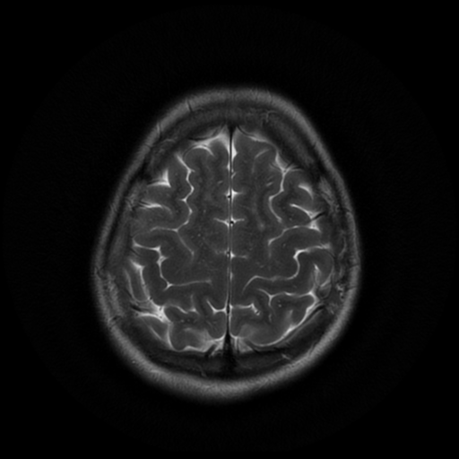
File:Cerebral autosomal dominant arteriopathy with subcortical infarcts and leukoencephalopathy (CADASIL) (Radiopaedia 41018-43768 Ax T2 PROP 17).png

From NC Commons
Jump to navigation Jump to search

Summary:

Description
  • Radiopaedia case ID: 41018
  • Image ID: 17364161
  • Image stack position: 17/20
  • Plane projection: Ax T2 PROP
  • Study findings: Moderately extensive foci of increased FLAIR and T2 signal evident within the periventricular white matter and subcortical white matter in the frontal and parietal lobes and sub insular white matter unchanged in comparison to the previous scan from two years prior. Small foci are evident within the basal ganglia and the largest confluent area is laterally on the right frontal lobe. Further FLAIR hyperintensity is evident in the subcortical white matter of the anterior temporal lobes, a more specific feature of CADASIL. There is no evidence of restricted diffusion. No critical intracranial vascular stenosis. Impression: Stable appearances and features suspicious for CADASIL.
  • Modality: MRI
  • System: Central Nervous System
  • Findings: Represented within the frontal and parietal lobe white matter bilaterally, in a fairly symmetrical distribution, numerous well circumscribed and irregularly marginated zones of T2 hyperintense signal are appreciated extending well up to the vertex, many being distributed within the watershed distribution of the frontal lobes bilaterally, and also at the capsular margins. Overall findings are well in keeping with areas of old subcortical and deep white matter infarcts/leuko-encephalopathy. None of these lesions demonstrate restricted diffusion on DWI sequence. No true periventricular white matter signal change is seen. No posterior fossa abnormality is seen. Sulcal, cistern and ventricular anatomy of the anterior and posterior circulation throughout. The right vertebral artery is dominant with normal junction point being demonstrated. Normal caliber of the basilar artery seen. The carotid syphons are normal bilaterally and no supraclinoid disease is seen. There is no evidence of AVM or aneurism. Conclusion: The findings are well in keeping with a diagnosis of cerebral autosomal dominant arteriopathy with subcortical infarcts and leukoencephalopathy (CADASIL). No acute infarct is seen on this occasion.
Date Published: 12th Nov 2015
Source https://radiopaedia.org/cases/cerebral-autosomal-dominant-arteriopathy-with-subcortical-infarcts-and-leukoencephalopathy-cadasil
Author Rajalakshmi Ramesh
Permission
(Permission-reusing-text)
http://creativecommons.org/licenses/by-nc-sa/3.0/

Licensing:

Attribution-NonCommercial-ShareAlike 3.0 Unported (CC BY-NC-SA 3.0)

File history

Click on a date/time to view the file as it appeared at that time.

Date/TimeThumbnailDimensionsUserComment
current10:52, 25 July 2021Thumbnail for version as of 10:52, 25 July 2021512 × 512 (163 KB) (talk | contribs)Radiopaedia project rID:41018 (batch #6898-57 C17)

There are no pages that use this file.

Metadata