File:Cerebral cavernoma with developmental venous anomaly (Radiopaedia 9218).jpg
Jump to navigation
Jump to search
Size of this preview: 534 × 599 pixels. Other resolutions: 214 × 240 pixels | 428 × 480 pixels | 738 × 828 pixels.
Original file (738 × 828 pixels, file size: 67 KB, MIME type: image/jpeg)
Summary:
- Radiopaedia case ID: 9218
- Image ID: 381943
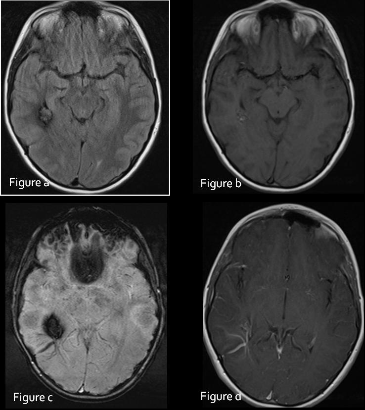
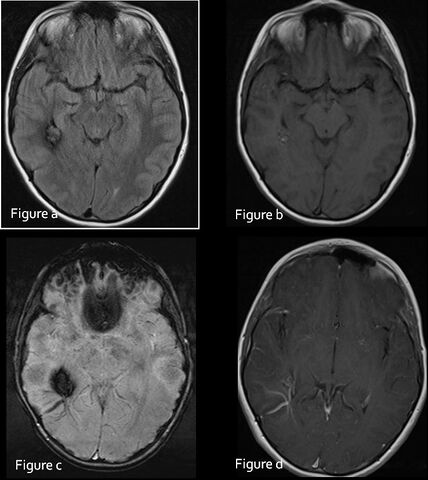
- Study findings: Figure a: axial FLAIR reveals a lobulated lesion with a low signal rim suggestive of hemosiderin in keeping with a cavernoma. A subtle flow void is seen extending to the cortical surface suggestive of an associated venous anomaly.Figure b: axial T1 reveals areas of increased signal within the lesion in keeping with methemoglobin suggestive of recent hemorrhage.Figure c: is an axial SWI image revealing the expected low signal appearance of a cavernoma due to the susceptibility effects of blood products. The venous architecture of DVA is much better visualized than on the other sequences.Figure d: is a contrast enhanced T1 image revealing the enhancement pattern of the venous anomaly. The carvernoma is less well visualized than on the SWI image
- Modality: MRI
- System: Central Nervous System
- Findings: Figure a: axial FLAIR reveals a lobulated lesion with a low signal rim suggestive of hemosiderin in keeping with a cavernoma. A subtle flow void is seen extending to the cortical surface suggestive of an associated venous anomaly. Figure b: axial T1 reveals areas of increased signal within the lesion in keeping with methemoglobin suggestive of recent hemorrhage. Figure c: is an axial SWI image revealing the expected low signal appearance of a cavernoma due to the susceptibility effects of blood products. The venous architecture of DVA is much better visualized than on the other sequences. Figure d: is a contrast enhanced T1 image revealing the enhancement pattern of the venous anomaly. The carvernoma is less well visualized than on the SWI image
- Published: 25th Mar 2010
- Source: https://radiopaedia.org/cases/cerebral-cavernoma-with-developmental-venous-anomaly
- Author: Sandeep Bhuta
- Permission: http://creativecommons.org/licenses/by-nc-sa/3.0/
Licensing:
Attribution-NonCommercial-ShareAlike 3.0 Unported (CC BY-NC-SA 3.0)
File history
Click on a date/time to view the file as it appeared at that time.
| Date/Time | Thumbnail | Dimensions | User | Comment | |
|---|---|---|---|---|---|
| current | 13:37, 20 March 2021 | | 738 × 828 (67 KB) | Fæ (talk | contribs) | Radiopaedia project rID:9218 (batch #6636) |
You cannot overwrite this file.
File usage
There are no pages that use this file.
.jpg&oldid=8859256)