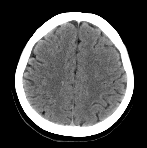
File:Cerebral cavernous venous malformations (Radiopaedia 48117-52945 Axial non-contrast 24).png
Jump to navigation
Jump to search
Cerebral_cavernous_venous_malformations_(Radiopaedia_48117-52945_Axial_non-contrast_24).png (512 × 515 pixels, file size: 55 KB, MIME type: image/png)
Summary:
| Description |
|
| Date | Published: 13th Oct 2016 |
| Source | https://radiopaedia.org/cases/cerebral-cavernous-venous-malformations |
| Author | Frank Gaillard |
| Permission (Permission-reusing-text) |
http://creativecommons.org/licenses/by-nc-sa/3.0/ |
Licensing:
Attribution-NonCommercial-ShareAlike 3.0 Unported (CC BY-NC-SA 3.0)
File history
Click on a date/time to view the file as it appeared at that time.
| Date/Time | Thumbnail | Dimensions | User | Comment | |
|---|---|---|---|---|---|
| current | 04:36, 26 July 2021 |  | 512 × 515 (55 KB) | Fæ (talk | contribs) | Radiopaedia project rID:48117 (batch #6929-24 A24) |
You cannot overwrite this file.
File usage
There are no pages that use this file.
