File:Cerebral embolic infarcts (embolic shower) (Radiopaedia 57395-64342 Axial DWI 5).png

From NC Commons
Jump to navigation Jump to search

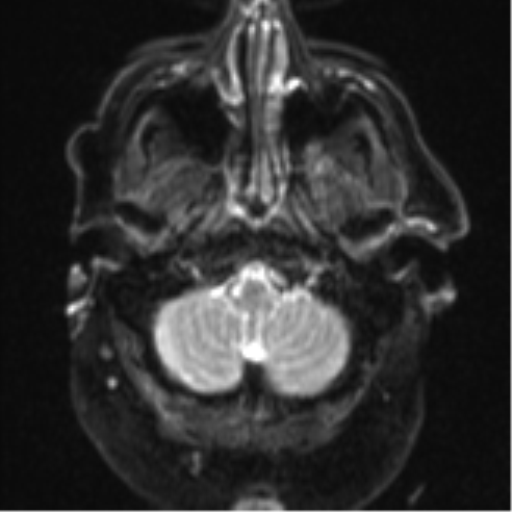
Cerebral_embolic_infarcts_(embolic_shower)_(Radiopaedia_57395-64342_Axial_DWI_5).png(512 × 512 pixels, file size: 88 KB, MIME type: image/png)

Summary:

Description
  • Radiopaedia case ID: 57395
  • Image ID: 34739612
  • Image stack position: 5/72
  • Plane projection: Axial
  • Aux modality: DWI
  • Study description: MRI Brain
  • Study findings: There are multiple small foci of diffusion restriction scattered throughout the brain parenchyma within all the major vascular territories, including the brainstem and cerebellar hemispheres. These demonstrate increased T2 and FLAIR signal and, although no definitive collection is appreciated within these small lesions, in this clinical scenario, septic emboli is the most favored diagnosis. The appearances are not of the suspected venous thrombosis. A small focus of susceptibility artefact in the right frontal lobe may represent micro-hemorrhage or a small cavernoma. No signs of an acute brain hemorrhage. The remainder of the brain, the ventricles, and basal cisterns appear unremarkable. MRA time-of-flight, although motion degraded, appears overall unremarkable when correlated to the previous CTA, with no evidence of acute abnormalities or convincing vasoconstriction (not shown). Conclusion: Embolic shower throughout the supra and infratentorial brain is likely septic in nature given the stated aortic root abscess. No convincing mycotic aneurysm within limitations of the motion degraded MRA.
  • Modality: MRI
  • System: Central Nervous System
  • Findings: There are new multiple rounded hypodensities of bilateral cerebral hemispheres involving the left centrum semiovale and bilateral superior frontal gyri. There is also a possible small hypodensity of the right cerebellar hemisphere. The previous tiny focus of subarachnoid blood of the right anterior frontal lobe has now resolved. No new focus of intracranial hemorrhage. No hydrocephalus or evidence of cerebral herniation. No suspicious bony lesion. Conclusion: Multiple bilateral white matter hypodensities, more prominent since the CT from 5 days prior, are suspicious for ischemic foci from septic emboli in this clinical setting. Small evolving abscesses are also possible, and MRI is recommended to further clarify. The previous tiny volume of right frontal subarachnoid blood has resolved. No hydrocephalus.
Date Published: 4th Feb 2018
Source https://radiopaedia.org/cases/cerebral-embolic-infarcts-embolic-shower-1
Author Bruno Di Muzio
Permission
(Permission-reusing-text)
http://creativecommons.org/licenses/by-nc-sa/3.0/

Licensing:

Attribution-NonCommercial-ShareAlike 3.0 Unported (CC BY-NC-SA 3.0)

File history

Click on a date/time to view the file as it appeared at that time.

Date/TimeThumbnailDimensionsUserComment
current16:38, 26 July 2021Thumbnail for version as of 16:38, 26 July 2021512 × 512 (88 KB) (talk | contribs)Radiopaedia project rID:57395 (batch #6949-5 A5)

Metadata