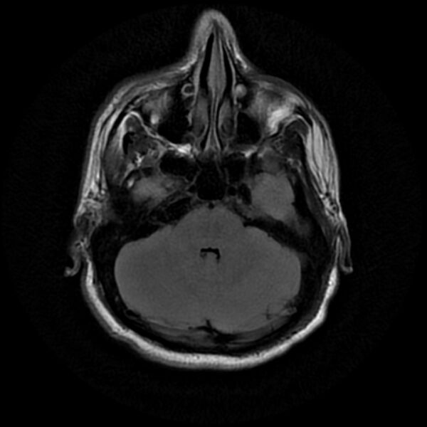
File:Cerebral fat embolism (Radiopaedia 37510-39363 Axial FLAIR 8).jpg
Jump to navigation
Jump to search

Size of this preview: 600 × 600 pixels. Other resolutions: 240 × 240 pixels | 630 × 630 pixels.
Original file (630 × 630 pixels, file size: 110 KB, MIME type: image/jpeg)
Summary:
| Description |
|
| Date | Published: 11th Jun 2015 |
| Source | https://radiopaedia.org/cases/cerebral-fat-embolism-4 |
| Author | Ahmed Abdrabou |
| Permission (Permission-reusing-text) |
http://creativecommons.org/licenses/by-nc-sa/3.0/ |
Licensing:
Attribution-NonCommercial-ShareAlike 3.0 Unported (CC BY-NC-SA 3.0)
File history
Click on a date/time to view the file as it appeared at that time.
| Date/Time | Thumbnail | Dimensions | User | Comment | |
|---|---|---|---|---|---|
| current | 19:23, 26 July 2021 |  | 630 × 630 (110 KB) | Fæ (talk | contribs) | Radiopaedia project rID:37510 (batch #6952-54 C8) |
You cannot overwrite this file.
File usage
There are no pages that use this file.