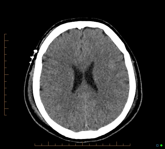
File:Cerebral fat embolism (Radiopaedia 85521-101224 Axial non-contrast 33).jpg
Jump to navigation
Jump to search
Cerebral_fat_embolism_(Radiopaedia_85521-101224_Axial_non-contrast_33).jpg (575 × 522 pixels, file size: 98 KB, MIME type: image/jpeg)
Summary:
| Description |
|
| Date | Published: 2nd Jan 2021 |
| Source | https://radiopaedia.org/cases/cerebral-fat-embolism-8 |
| Author | James Harvey |
| Permission (Permission-reusing-text) |
http://creativecommons.org/licenses/by-nc-sa/3.0/ |
Licensing:
Attribution-NonCommercial-ShareAlike 3.0 Unported (CC BY-NC-SA 3.0)
File history
Click on a date/time to view the file as it appeared at that time.
| Date/Time | Thumbnail | Dimensions | User | Comment | |
|---|---|---|---|---|---|
| current | 22:22, 26 July 2021 |  | 575 × 522 (98 KB) | Fæ (talk | contribs) | Radiopaedia project rID:85521 (batch #6957-33 A33) |
You cannot overwrite this file.
File usage
The following page uses this file:
