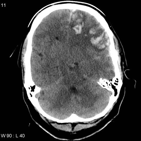
File:Cerebral hemorrhagic contusion with subdural and subarachnoid hemorrhage (Radiopaedia 10680-19197 Axial non-contrast 7).jpg
Jump to navigation
Jump to search

Size of this preview: 601 × 599 pixels. Other resolutions: 241 × 240 pixels | 481 × 480 pixels | 766 × 764 pixels.
Original file (766 × 764 pixels, file size: 85 KB, MIME type: image/jpeg)
Summary:
| Description |
|
| Date | Published: 5th Sep 2010 |
| Source | https://radiopaedia.org/cases/cerebral-haemorrhagic-contusion-with-subdural-and-subarachnoid-haemorrhage |
| Author | Frank Gaillard |
| Permission (Permission-reusing-text) |
http://creativecommons.org/licenses/by-nc-sa/3.0/ |
Licensing:
Attribution-NonCommercial-ShareAlike 3.0 Unported (CC BY-NC-SA 3.0)
File history
Click on a date/time to view the file as it appeared at that time.
| Date/Time | Thumbnail | Dimensions | User | Comment | |
|---|---|---|---|---|---|
| current | 19:51, 27 July 2021 | | 766 × 764 (85 KB) | Fæ (talk | contribs) | Radiopaedia project rID:10680 (batch #6978-7 A7) |
You cannot overwrite this file.
File usage
There are no pages that use this file.
.jpg&oldid=830234)