File:Cerebral hemorrhagic contusions- temporal evolution (Radiopaedia 40224-42755 AXIAL THICK 4).png

From NC Commons
Jump to navigation Jump to search

Cerebral_hemorrhagic_contusions-_temporal_evolution_(Radiopaedia_40224-42755_AXIAL_THICK_4).png(512 × 512 pixels, file size: 96 KB, MIME type: image/png)

Summary:

Description
  • Radiopaedia case ID: 40224
  • Image ID: 16530398
  • Image stack position: 4/36
  • Plane projection: AXIAL THICK
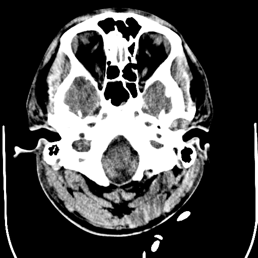
  • Study description: CT Brain - Day 7
  • Study findings: Bifrontal craniectomy performed in the interval since the previous scan. Bifrontal scalp high density which may represent surgical material or hematoma. Right frontal EVD tip abuts the right foramen of Monro. The extensive right frontal and anterior temporal contusions are unchanged in size with slightly more surrounding hypodensity on today's study. Degree of midline shift to the left is approximately 10 mm (previously 8mm). Left cerebellar hemisphere contusion unchanged. Surface CSF spaces are moderately more effaced than on the previous study. Ventricular size is unchanged, with the right ventricle being more effaced than the left. The basal cisterns the patent. Conclusion: Overally, marginal increase in midline shift and diffuse sulcal effacement since the previous study. No new hemorrhage or infarction.​
  • Modality: CT
  • System: Central Nervous System
  • Findings: Bilateral frontal contusions, worse on the right. Subarachnoid hemorrhage over the right frontal lobe are noted in the sylvian fissures bilaterally. There is midline shift to the left of 3 mm. The basal cisterns are effaced. Left posterior fossa hemorrhage is probably extra-axial. Bilateral subtle skull base bone fractures which do not extend to the occipital condyles. On the right, the occipital fracture extends into the right jugular foramen through the vestibular apparatus across the facial nerve canal and in the right temporomandibular joint. The fracture also involves the right carotid canal. There are locules of gas seen within the semi circular canals. Blood fills the right middle ear cleft.
Date Published: 20th Oct 2015
Source https://radiopaedia.org/cases/cerebral-haemorrhagic-contusions-temporal-evolution
Author Bruno Di Muzio
Permission
(Permission-reusing-text)
http://creativecommons.org/licenses/by-nc-sa/3.0/

Licensing:

Attribution-NonCommercial-ShareAlike 3.0 Unported (CC BY-NC-SA 3.0)

File history

Click on a date/time to view the file as it appeared at that time.

Date/TimeThumbnailDimensionsUserComment
current18:41, 27 July 2021Thumbnail for version as of 18:41, 27 July 2021512 × 512 (96 KB) (talk | contribs)Radiopaedia project rID:40224 (batch #6977-4 A4)

Metadata