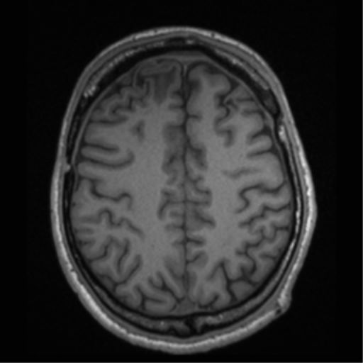
File:Cerebral hemorrhagic contusions- temporal evolution (Radiopaedia 40224-42757 Axial T1 57).png

From NC Commons
Jump to navigation Jump to search

Cerebral_hemorrhagic_contusions-_temporal_evolution_(Radiopaedia_40224-42757_Axial_T1_57).png(512 × 512 pixels, file size: 183 KB, MIME type: image/png)

Summary:

Description
  • Radiopaedia case ID: 40224
  • Image ID: 16532753
  • Image stack position: 57/79
  • Plane projection: Axial
  • Aux modality: T1
  • Study description: MRI Brain - Epilepsy protocol (3 years later)
  • Study findings: No previous MRIs available for comparison. Review is made with the preceding CT scans. Previous bifrontal craniectomy and previous bilateral drain tubes placement is noted. Extensive regions of encephalomalacia are present at the sites of the previous shearing injuries in both frontal lobes. This involves the anterior and inferior aspect of the right frontal lobe with less severe encephalomalacia involving the anterior aspect of the left frontal lobe as well as around the line of the previously placed left drain tube. Less severe changes affect the right temporal lobe. A small focus of signal change is noted within the left thalamus. Further regions of encephalomalacia and hemosiderin staining are noted within the left cerebellar hemisphere with extension into the vermis. Volume loss companies the regions of encephalomalacia and is reflected in volume loss within the anterior aspect of the corpus callosum. No abnormality of the parietal lobes is identified. The hippocampi remain of good volume Conclusion: Previous severe head trauma. Extensive bifrontal regions of encephalomalacia with further involvement of the temporal lobes and left cerebellar hemisphere. No abnormality of the parietal lobe is identified​
  • Modality: MRI
  • System: Central Nervous System
  • Findings: Bilateral frontal contusions, worse on the right. Subarachnoid hemorrhage over the right frontal lobe are noted in the sylvian fissures bilaterally. There is midline shift to the left of 3 mm. The basal cisterns are effaced. Left posterior fossa hemorrhage is probably extra-axial. Bilateral subtle skull base bone fractures which do not extend to the occipital condyles. On the right, the occipital fracture extends into the right jugular foramen through the vestibular apparatus across the facial nerve canal and in the right temporomandibular joint. The fracture also involves the right carotid canal. There are locules of gas seen within the semi circular canals. Blood fills the right middle ear cleft.
Date Published: 20th Oct 2015
Source https://radiopaedia.org/cases/cerebral-haemorrhagic-contusions-temporal-evolution
Author Bruno Di Muzio
Permission
(Permission-reusing-text)
http://creativecommons.org/licenses/by-nc-sa/3.0/

Licensing:

Attribution-NonCommercial-ShareAlike 3.0 Unported (CC BY-NC-SA 3.0)

File history

Click on a date/time to view the file as it appeared at that time.

Date/TimeThumbnailDimensionsUserComment
current19:25, 27 July 2021Thumbnail for version as of 19:25, 27 July 2021512 × 512 (183 KB) (talk | contribs)Radiopaedia project rID:40224 (batch #6977-233 E57)

Metadata