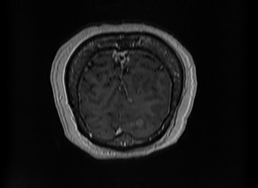
File:Cerebral metastases from lung cancer with amyloid angiopathy and cerebellopontine angle meningioma (Radiopaedia 74306-85191 Coronal T1 C+ 57).jpg
Jump to navigation
Jump to search
No higher resolution available.
Cerebral_metastases_from_lung_cancer_with_amyloid_angiopathy_and_cerebellopontine_angle_meningioma_(Radiopaedia_74306-85191_Coronal_T1_C+_57).jpg (512 × 373 pixels, file size: 47 KB, MIME type: image/jpeg)
Summary:
| Description |
|
| Date | Published: 27th Feb 2020 |
| Source | https://radiopaedia.org/cases/cerebral-metastases-from-lung-cancer-with-amyloid-angiopathy-and-cerebellopontine-angle-meningioma |
| Author | Dr Ammar Haouimi |
| Permission (Permission-reusing-text) |
http://creativecommons.org/licenses/by-nc-sa/3.0/ |
Licensing:
Attribution-NonCommercial-ShareAlike 3.0 Unported (CC BY-NC-SA 3.0)
File history
Click on a date/time to view the file as it appeared at that time.
| Date/Time | Thumbnail | Dimensions | User | Comment | |
|---|---|---|---|---|---|
| current | 06:24, 28 July 2021 | | 512 × 373 (47 KB) | Fæ (talk | contribs) | Radiopaedia project rID:74306 (batch #7006-202 G57) |
You cannot overwrite this file.
File usage
The following page uses this file:
.jpg&oldid=834357)