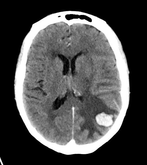
File:Cerebral metastasis (Radiopaedia 46744-51260 Axial C+ delayed 17).png
Jump to navigation
Jump to search
Cerebral_metastasis_(Radiopaedia_46744-51260_Axial_C+_delayed_17).png (512 × 574 pixels, file size: 59 KB, MIME type: image/png)
Summary:
| Description |
|
| Date | Published: 15th Jul 2016 |
| Source | https://radiopaedia.org/cases/cerebral-metastasis-3 |
| Author | Henry Knipe |
| Permission (Permission-reusing-text) |
http://creativecommons.org/licenses/by-nc-sa/3.0/ |
Licensing:
Attribution-NonCommercial-ShareAlike 3.0 Unported (CC BY-NC-SA 3.0)
File history
Click on a date/time to view the file as it appeared at that time.
| Date/Time | Thumbnail | Dimensions | User | Comment | |
|---|---|---|---|---|---|
| current | 13:23, 28 July 2021 |  | 512 × 574 (59 KB) | Fæ (talk | contribs) | Radiopaedia project rID:46744 (batch #7023-17 A17) |
You cannot overwrite this file.
File usage
There are no pages that use this file.
