File:Cerebral metastasis (Radiopaedia 80088).jpg

From NC Commons
Jump to navigation Jump to search

Cerebral_metastasis_(Radiopaedia_80088).jpg(512 × 512 pixels, file size: 18 KB, MIME type: image/jpeg)

Summary:

Description
  • Radiopaedia case ID: 80088
  • Image ID: 53123525
  • Modality: CT
  • System: Oncology
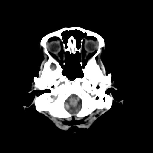
  • Findings: There is a fairly well circumscribed lobulated hyperdense intra-axial space occupying lesion in left area centrum semiovale extending towards corona radiata creeping along the body of left lateral ventricle reaching upto the left basal ganglia and thalamus with suggestion of possible subependymal infiltration in body of the left lateral ventricle. This is measuring approximately 4.0 x 2.3 cm in AP and TR dimensions. There is associated significant disproportionate vasogenic edema in left cerebral hemisphere including basal ganglia and thalamus, causing mass effect resulting in effacement of the left lateral ventricle and mild contralateral midline shift of 0.3 cm. Another focal area of vasogenic edema is present in right corona radiata. In keeping with history of treated carcinoma of the breast, imaging features are highly suggestive of brain metastasis. Other differential would include cerebral abscess. Contrast enhanced MRI is recommended for further evaluation.
Date Published: 21st Jul 2020
Source https://radiopaedia.org/cases/cerebral-metastasis-7
Author Muhammad Asadullah Munir
Permission
(Permission-reusing-text)
http://creativecommons.org/licenses/by-nc-sa/3.0/

Licensing:

Attribution-NonCommercial-ShareAlike 3.0 Unported (CC BY-NC-SA 3.0)

File history

Click on a date/time to view the file as it appeared at that time.

Date/TimeThumbnailDimensionsUserComment
current13:59, 28 July 2021Thumbnail for version as of 13:59, 28 July 2021512 × 512 (18 KB) (talk | contribs)Radiopaedia project rID:80088 (batch #7024)