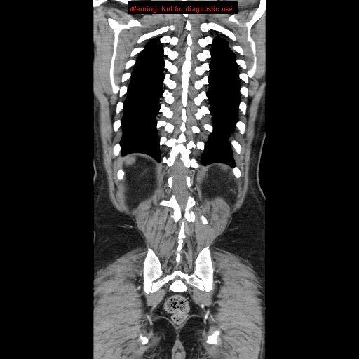
File:Cerebral metastasis - colorectal carcinoma (Radiopaedia 8581-83548 A 69).jpg
Jump to navigation
Jump to search
Cerebral_metastasis_-_colorectal_carcinoma_(Radiopaedia_8581-83548_A_69).jpg (512 × 512 pixels, file size: 107 KB, MIME type: image/jpeg)
Summary:
| Description |
|
| Date | Published: 14th Feb 2010 |
| Source | https://radiopaedia.org/cases/cerebral-metastasis-colorectal-carcinoma |
| Author | Hani Makky Al Salam |
| Permission (Permission-reusing-text) |
http://creativecommons.org/licenses/by-nc-sa/3.0/ |
Licensing:
Attribution-NonCommercial-ShareAlike 3.0 Unported (CC BY-NC-SA 3.0)
File history
Click on a date/time to view the file as it appeared at that time.
| Date/Time | Thumbnail | Dimensions | User | Comment | |
|---|---|---|---|---|---|
| current | 15:40, 28 July 2021 |  | 512 × 512 (107 KB) | Fæ (talk | contribs) | Radiopaedia project rID:8581 (batch #7030-69 A69) |
You cannot overwrite this file.
File usage
The following page uses this file:
