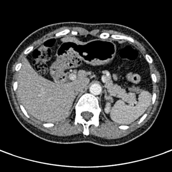
File:Cerebral ring enhancing lesions - cerebral metastases (Radiopaedia 44922-48819 A 279).jpg
Jump to navigation
Jump to search
Cerebral_ring_enhancing_lesions_-_cerebral_metastases_(Radiopaedia_44922-48819_A_279).jpg (594 × 594 pixels, file size: 94 KB, MIME type: image/jpeg)
Summary:
| Description |
|
| Date | Published: 21st May 2016 |
| Source | https://radiopaedia.org/cases/cerebral-ring-enhancing-lesions-cerebral-metastases |
| Author | Ian Bickle |
| Permission (Permission-reusing-text) |
http://creativecommons.org/licenses/by-nc-sa/3.0/ |
Licensing:
Attribution-NonCommercial-ShareAlike 3.0 Unported (CC BY-NC-SA 3.0)
File history
Click on a date/time to view the file as it appeared at that time.
| Date/Time | Thumbnail | Dimensions | User | Comment | |
|---|---|---|---|---|---|
| current | 18:34, 29 July 2021 |  | 594 × 594 (94 KB) | Fæ (talk | contribs) | Radiopaedia project rID:44922 (batch #7057-279 A279) |
You cannot overwrite this file.
File usage
The following page uses this file:
