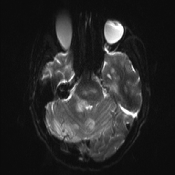
File:Cerebral toxoplasmosis (Radiopaedia 53993-60132 Axial DWI 8).jpg

From NC Commons
Jump to navigation Jump to search

Cerebral_toxoplasmosis_(Radiopaedia_53993-60132_Axial_DWI_8).jpg(588 × 588 pixels, file size: 58 KB, MIME type: image/jpeg)

Summary:

Description
  • Radiopaedia case ID: 53993
  • Image ID: 30986908
  • Image stack position: 8/54
  • Plane projection: Axial
  • Aux modality: DWI
  • Study findings: Multiple lesions of variable size measuring from 9 to 25 mm with surrounding edema throughout the brain including, cerebral hemispheres, cerebellum and brain stem. The lesions are hyperintense on T2 and involving the subcortical regions, white matter and basal ganglia. The largest one is in the left basal ganglia. Several have low T2 signal rims. Focal areas of restricted diffusion in a few lesions in the parietal lobe. No intralesional calcification. Variable enhancement with faint ring enhancement of a minority of the lesions. Normal paranasal sinuses. Mastoids are normal and the calvarium demonstrates no abnormality.
  • Modality: MRI
  • System: Central Nervous System
  • Findings: Ill defined non-enhancing low attenuation in the left lentiform nucleus and internal capsule. Similar ill defined area of low attenuation in the left posterior parietal lobe, including the post central gyrus. No abnormal enhancement.
Date Published: 30th Aug 2017
Source https://radiopaedia.org/cases/cerebral-toxoplasmosis-11
Author Ian Bickle
Permission
(Permission-reusing-text)
http://creativecommons.org/licenses/by-nc-sa/3.0/

Licensing:

Attribution-NonCommercial-ShareAlike 3.0 Unported (CC BY-NC-SA 3.0)

File history

Click on a date/time to view the file as it appeared at that time.

Date/TimeThumbnailDimensionsUserComment
current20:44, 29 July 2021Thumbnail for version as of 20:44, 29 July 2021588 × 588 (58 KB) (talk | contribs)Radiopaedia project rID:53993 (batch #7061-117 E8)