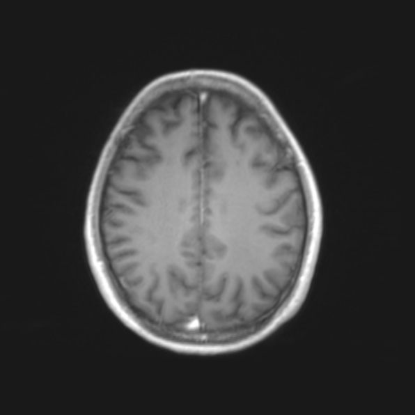
File:Cerebral toxoplasmosis (Radiopaedia 53993-61435 Axial T1 17).jpg
Jump to navigation
Jump to search
Cerebral_toxoplasmosis_(Radiopaedia_53993-61435_Axial_T1_17).jpg (596 × 596 pixels, file size: 39 KB, MIME type: image/jpeg)
Summary:
| Description |
|
| Date | Published: 30th Aug 2017 |
| Source | https://radiopaedia.org/cases/cerebral-toxoplasmosis-11 |
| Author | Ian Bickle |
| Permission (Permission-reusing-text) |
http://creativecommons.org/licenses/by-nc-sa/3.0/ |
Licensing:
Attribution-NonCommercial-ShareAlike 3.0 Unported (CC BY-NC-SA 3.0)
File history
Click on a date/time to view the file as it appeared at that time.
| Date/Time | Thumbnail | Dimensions | User | Comment | |
|---|---|---|---|---|---|
| current | 21:01, 29 July 2021 |  | 596 × 596 (39 KB) | Fæ (talk | contribs) | Radiopaedia project rID:53993 (batch #7061-17 A17) |
You cannot overwrite this file.
File usage
There are no pages that use this file.
