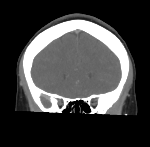
File:Cerebral venous infarct related to dural venous sinus thromboses (Radiopaedia 35292-36804 Coronal C+ delayed 19).png
Jump to navigation
Jump to search
No higher resolution available.
Cerebral_venous_infarct_related_to_dural_venous_sinus_thromboses_(Radiopaedia_35292-36804_Coronal_C+_delayed_19).png (521 × 512 pixels, file size: 96 KB, MIME type: image/png)
Summary:
| Description |
|
| Date | Published: 5th Apr 2015 |
| Source | https://radiopaedia.org/cases/cerebral-venous-infarct-related-to-dural-venous-sinus-thromboses |
| Author | Bruno Di Muzio |
| Permission (Permission-reusing-text) |
http://creativecommons.org/licenses/by-nc-sa/3.0/ |
Licensing:
Attribution-NonCommercial-ShareAlike 3.0 Unported (CC BY-NC-SA 3.0)
File history
Click on a date/time to view the file as it appeared at that time.
| Date/Time | Thumbnail | Dimensions | User | Comment | |
|---|---|---|---|---|---|
| current | 21:14, 30 July 2021 | | 521 × 512 (96 KB) | Fæ (talk | contribs) | Radiopaedia project rID:35292 (batch #7105-167 E19) |
You cannot overwrite this file.
File usage
The following 2 pages use this file:
.png&oldid=855428)