File:Cerebral venous infarction - hemorrhagic (Radiopaedia 81625-95505 Sagittal MRV 63).jpg
Jump to navigation
Jump to search
No higher resolution available.
Cerebral_venous_infarction_-_hemorrhagic_(Radiopaedia_81625-95505_Sagittal_MRV_63).jpg (217 × 217 pixels, file size: 16 KB, MIME type: image/jpeg)
Summary:
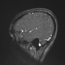
| Description |
|
| Date | Published: 24th Nov 2020 |
| Source | https://radiopaedia.org/cases/cerebral-venous-infarction-hemorrhagic |
| Author | Mostafa El-Feky |
| Permission (Permission-reusing-text) |
http://creativecommons.org/licenses/by-nc-sa/3.0/ |
Licensing:
Attribution-NonCommercial-ShareAlike 3.0 Unported (CC BY-NC-SA 3.0)
File history
Click on a date/time to view the file as it appeared at that time.
| Date/Time | Thumbnail | Dimensions | User | Comment | |
|---|---|---|---|---|---|
| current | 18:46, 30 July 2021 | | 217 × 217 (16 KB) | Fæ (talk | contribs) | Radiopaedia project rID:81625 (batch #7103-246 I63) |
You cannot overwrite this file.
File usage
The following page uses this file:
.jpg&oldid=854422)