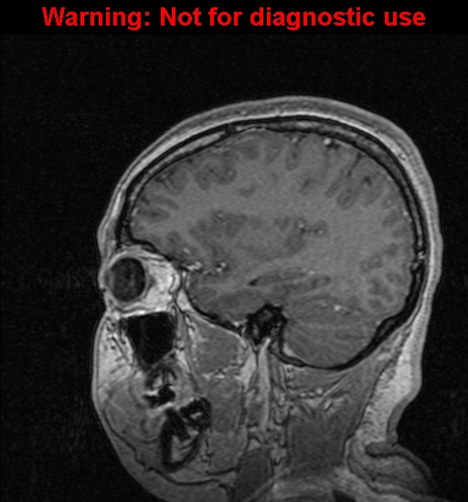
File:Cerebral venous thrombosis (Radiopaedia 37224-39208 Sagittal T1 C+ 44).jpg
Jump to navigation
Jump to search
No higher resolution available.
Cerebral_venous_thrombosis_(Radiopaedia_37224-39208_Sagittal_T1_C+_44).jpg (512 × 549 pixels, file size: 135 KB, MIME type: image/jpeg)
Summary:
| Description |
|
| Date | Published: 22nd Jun 2015 |
| Source | https://radiopaedia.org/cases/cerebral-venous-thrombosis-5 |
| Author | Foroogh Jafari Mousavi |
| Permission (Permission-reusing-text) |
http://creativecommons.org/licenses/by-nc-sa/3.0/ |
Licensing:
Attribution-NonCommercial-ShareAlike 3.0 Unported (CC BY-NC-SA 3.0)
File history
Click on a date/time to view the file as it appeared at that time.
| Date/Time | Thumbnail | Dimensions | User | Comment | |
|---|---|---|---|---|---|
| current | 03:52, 31 July 2021 | | 512 × 549 (135 KB) | Fæ (talk | contribs) | Radiopaedia project rID:37224 (batch #7116-64 B44) |
You cannot overwrite this file.
File usage
The following page uses this file:
.jpg&oldid=858049)