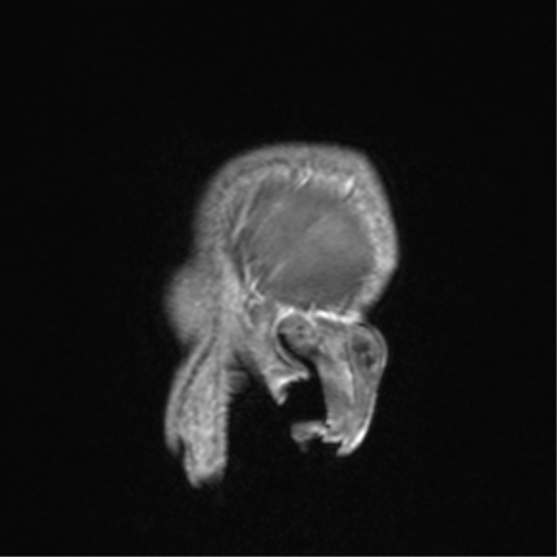
File:Cerebral venous thrombosis (Radiopaedia 38392-40469 Sagittal T1 C+ 84).png

From NC Commons
Jump to navigation Jump to search

Cerebral_venous_thrombosis_(Radiopaedia_38392-40469_Sagittal_T1_C+_84).png(512 × 512 pixels, file size: 143 KB, MIME type: image/png)

Summary:

Description
  • Radiopaedia case ID: 38392
  • Image ID: 14604244
  • Image stack position: 84/88
  • Plane projection: Sagittal
  • Aux modality: T1 C+
  • Study description: MRI Brain / MRV (follow-up 6 months later)
  • Study findings: Technique: Multiplanar, multisequence imaging has been obtained through the brain including post contrast images and MRV. Findings: No intra or extra-axial abnormality, mass, or collection. Ventricles and basal cisterns are normal in appearance. No abnormal enhancement. Cerebral venous sinuses, deep and superficial cerebral veins appear normal, with no signs of new venous thrombosis, although the caliber of the anterior two thirds of the superior sagittal sinus remains reduced. Flow is demonstrated in this region on phase contrast MRV. Within the left frontal lobe are numerous prominent transmedullary veins which drain into the internal cerebral veins on the left. These presumably represent enlarged collateral drainage pathways, and no abnormal arteries are demonstrated in the region (middle cerebral, middle meningeal or superficial temporal arteries) to suggest the presence of a dural arteriovenous venous fistula, however continued followup is recommended. On future imaging a MR angiogram would be of benefit.
  • Modality: MRI
  • System: Central Nervous System
  • Findings: There is a small volume of left frontal subarachnoid blood. The overlying left frontal superficial cortical veins and anterior superior sagittal sinus appear hyperdense. No definite intra-axial hemorrhage (possible small petechial component). Grey-white matter differentiation is preserved. No midline shift. No hydrocephalus. No skull fracture or subgaleal hematoma. Conclusion: Left frontal subarachnoid hemorrhage with findings suggestive of left frontal cortical vein thrombosis with involvement of the frontal aspect of the sagittal sinus. Whilst this may be an isolated finding an underlying dural AVF warrants exclusion. Emergent evaluation with CTV/CTA COW recommended in the first instance. CT Venogram There is a filling defect within the anterior superior sagittal sinus and hypoenhancement and additional filling defects in the left frontal superficial cortical veins. The posterior superior sagittal sinus and remainder of the venous sinuses opacify normally. CTA COW (not shown) The aortic arch has not been imaged. The right vertebral artery is dominant; the left arises from the arch. The left PCA has a fetal origin. The extracranial vessels, circle of Willis and vertebrobasilar system opacify normally. No evidence of an aneurysm, vascular malformation, dissection or significant stenosis. Conclusion: Findings are consistent with anterior superior sagittal sinus/left frontal superficial cortical vein thrombosis. No underlying dural AVF identified.
Date Published: 19th Jul 2015
Source https://radiopaedia.org/cases/cerebral-venous-thrombosis-6
Author Bruno Di Muzio
Permission
(Permission-reusing-text)
http://creativecommons.org/licenses/by-nc-sa/3.0/

Licensing:

Attribution-NonCommercial-ShareAlike 3.0 Unported (CC BY-NC-SA 3.0)

File history

Click on a date/time to view the file as it appeared at that time.

Date/TimeThumbnailDimensionsUserComment
current05:08, 31 July 2021Thumbnail for version as of 05:08, 31 July 2021512 × 512 (143 KB) (talk | contribs)Radiopaedia project rID:38392 (batch #7120-107 B84)

Metadata