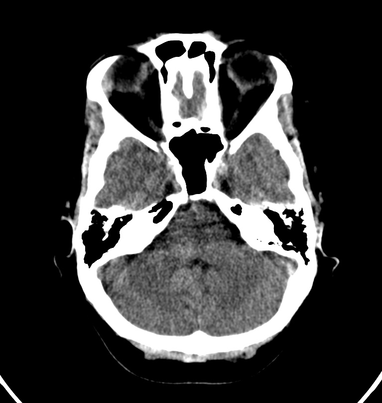
File:Cerebral venous thrombosis - CT only (Radiopaedia 41031-43778 Axial non-contrast 66).jpg
Jump to navigation
Jump to search
Size of this preview: 569 × 599 pixels. Other resolutions: 228 × 240 pixels | 456 × 480 pixels | 767 × 808 pixels.
Original file (767 × 808 pixels, file size: 100 KB, MIME type: image/jpeg)
Summary:
| Description |
|
| Date | Published: 18th Nov 2015 |
| Source | https://radiopaedia.org/cases/cerebral-venous-thrombosis-ct-only |
| Author | Bruno Di Muzio |
| Permission (Permission-reusing-text) |
http://creativecommons.org/licenses/by-nc-sa/3.0/ |
Licensing:
Attribution-NonCommercial-ShareAlike 3.0 Unported (CC BY-NC-SA 3.0)
File history
Click on a date/time to view the file as it appeared at that time.
| Date/Time | Thumbnail | Dimensions | User | Comment | |
|---|---|---|---|---|---|
| current | 05:42, 31 July 2021 | | 767 × 808 (100 KB) | Fæ (talk | contribs) | Radiopaedia project rID:41031 (batch #7121-66 A66) |
You cannot overwrite this file.
File usage
The following page uses this file:
.jpg&oldid=858778)