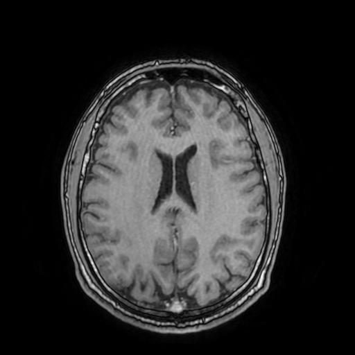
File:Cerebral venous thrombosis with secondary intracranial hypertension (Radiopaedia 89842-106957 Axial T1 C+ 113).jpg
Jump to navigation
Jump to search
Cerebral_venous_thrombosis_with_secondary_intracranial_hypertension_(Radiopaedia_89842-106957_Axial_T1_C+_113).jpg (512 × 512 pixels, file size: 22 KB, MIME type: image/jpeg)
Summary:
| Description |
|
| Date | Published: 24th May 2021 |
| Source | https://radiopaedia.org/cases/cerebral-venous-thrombosis-with-secondary-intracranial-hypertension-1 |
| Author | Dr Ammar Haouimi |
| Permission (Permission-reusing-text) |
http://creativecommons.org/licenses/by-nc-sa/3.0/ |
Licensing:
Attribution-NonCommercial-ShareAlike 3.0 Unported (CC BY-NC-SA 3.0)
File history
Click on a date/time to view the file as it appeared at that time.
| Date/Time | Thumbnail | Dimensions | User | Comment | |
|---|---|---|---|---|---|
| current | 12:31, 31 July 2021 |  | 512 × 512 (22 KB) | Fæ (talk | contribs) | Radiopaedia project rID:89842 (batch #7130-457 I113) |
You cannot overwrite this file.
File usage
The following page uses this file:
