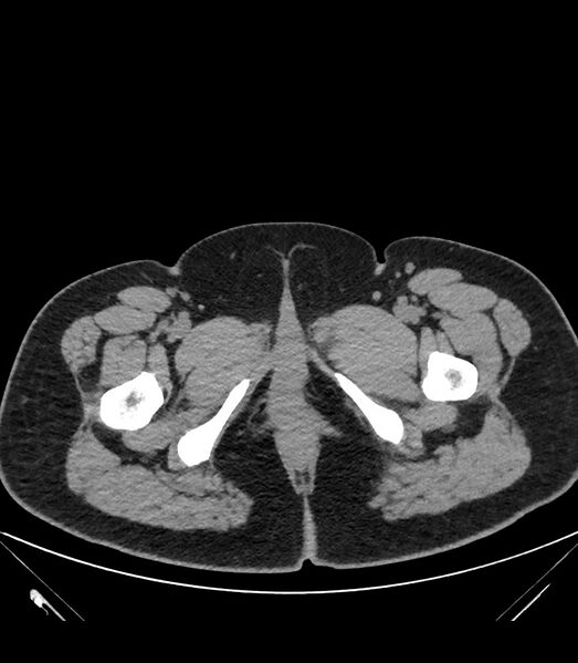
File:Cervical aortic arch with coarctation and aneurysms (Radiopaedia 44035-47552 Axial non-contrast 103).jpg

From NC Commons
Jump to navigation Jump to search

Original file(810 × 928 pixels, file size: 96 KB, MIME type: image/jpeg)

Summary:

Description
  • Radiopaedia case ID: 44035
  • Image ID: 21002303
  • Image stack position: 103/122
  • Plane projection: Axial
  • Aux modality: non-contrast
  • Study findings: The aorta arises normally from the left ventricle. Left-sided aortic arch and descending aorta are demonstrated. The aorta gives off three branches in the following order: innominate artery (brachiocephalic trunk), left common carotid artery and left subclavian artery. The segment of the aortic arch between the left common carotid artery and left subclavian artery is elongated and markedly tortuous with its apex positioned cephalad slightly above the level of the medial ends of the clavicles consistent with cervical aortic arch. This segment of the aortic arch also shows multiple saccular aneurysms. The largest saccular aneurysm exhibits wall calcifications. There is stenosis (coarctation) of the aortic arch after the take-off of the left common carotid artery. The descending segment of the thoracic aorta is smaller in caliber than usual suggestive of mild hypoplasia. The right internal thoracic artery is prominent and connects to the right external iliac artery via the superior and inferior epigastric arteries likely serving as a collateral. The left brachiocephalic vein courses behind the ascending segment of the aorta before it joins the superior vena cava.
  • Modality: CT
  • System: Vascular
  • Findings: This anteroposterior chest radiograph shows a widened mediastinum with a convex soft tissue overlying the left hilum.  Calcifications are seen along the margin of the convex soft tissue density.  A soft tissue density is also observed along the left paratracheal region. The transverse diameter of the heart is increased which may still be attributed to magnification. Apparent haziness in both lower lungs are possibly due to partial volume loss secondary to lack of deep inspiration.
Date Published: 9th Apr 2016
Source https://radiopaedia.org/cases/cervical-aortic-arch-with-coarctation-and-aneurysms
Author Vincent Tatco
Permission
(Permission-reusing-text)
http://creativecommons.org/licenses/by-nc-sa/3.0/

Licensing:

Attribution-NonCommercial-ShareAlike 3.0 Unported (CC BY-NC-SA 3.0)

File history

Click on a date/time to view the file as it appeared at that time.

Date/TimeThumbnailDimensionsUserComment
current16:08, 31 July 2021Thumbnail for version as of 16:08, 31 July 2021810 × 928 (96 KB) (talk | contribs)Radiopaedia project rID:44035 (batch #7141-103 A103)