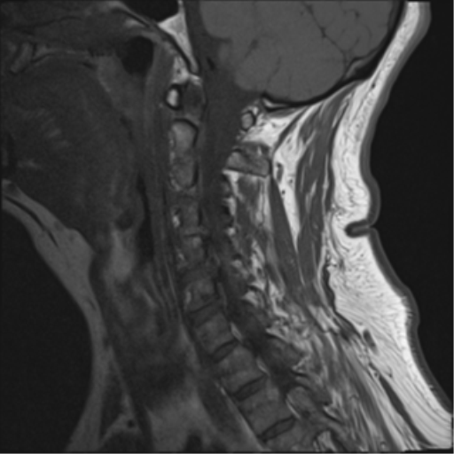
File:Cervical canal stenosis - OPLL and osteophytes (Radiopaedia 47329-51911 Sagittal T1 5).png

From NC Commons
Jump to navigation Jump to search

Cervical_canal_stenosis_-_OPLL_and_osteophytes_(Radiopaedia_47329-51911_Sagittal_T1_5).png(512 × 512 pixels, file size: 118 KB, MIME type: image/png)

Summary:

Description
  • Radiopaedia case ID: 47329
  • Image ID: 24535476
  • Image stack position: 5/15
  • Plane projection: Sagittal
  • Aux modality: T1
  • Study description: MRI Cervical spine
  • Study findings: The vertebral bodies have normal height and alignment. Nonunion of the C1 posterior arch on the right (more conspicuous on CT) is consistent with an anatomical variant. There is degenerative ankylosis of C4 and C5 vertebral bodies associated with MODIC 2 signal changes of their interposed endplates. No bone marrow edema, facet joint effusion or abnormal disc signal. Anterior to the C4-C6 vertebral body as well as a thin layer of prevertebral high signal, however, this is not associated with evidence of anterior longitudinal ligament or disc disruption. The posterior longitudinal ligament and ligamentum flavum appear intact. No posterior soft tissue bruising or hematoma. No epidural abscess. Bulky osteophytes/ossification of the posterior longitudinal ligament from C4 to C6/7 is eccentric to the right. This causes severe canal stenosis and cord compression posterior to C5 and C5/6; associated cord edema extends between C4/5 and C6/7. Disc osteophyte complex at C4/5 impinges upon the passing right ventral nerve root and causes moderate right and mild left foraminal stenosis. At C5-6 the eccentricOPLL/osteophytosis impinges the pre-foraminal right C6 nerve root and left uncovertebral osteophytes to cause moderate left foraminal narrowing. At C6-7, there is a mild indentation of the thecal sac by right paracentral protrusion without cord contact or foraminal stenosis.
  • Modality: MRI
  • System: Spine
  • Findings: No acute cervical spine injury. Non-union of the posterior arch on the right of C1 which is long-standing, either an old fracture or congenital abnormality. Large posterior disc osteophyte complex at C5-6 causes cord compression and spinal canal stenosis. Given the degree of cord compression, neurological assessment and possible elective outpatient MRI may be indicated to further assess.
Date Published: 16th Jul 2017
Source https://radiopaedia.org/cases/cervical-canal-stenosis-opll-and-osteophytes
Author Bruno Di Muzio
Permission
(Permission-reusing-text)
http://creativecommons.org/licenses/by-nc-sa/3.0/

Licensing:

Attribution-NonCommercial-ShareAlike 3.0 Unported (CC BY-NC-SA 3.0)

File history

Click on a date/time to view the file as it appeared at that time.

Date/TimeThumbnailDimensionsUserComment
current18:54, 31 July 2021Thumbnail for version as of 18:54, 31 July 2021512 × 512 (118 KB) (talk | contribs)Radiopaedia project rID:47329 (batch #7146-20 B5)

There are no pages that use this file.

Metadata