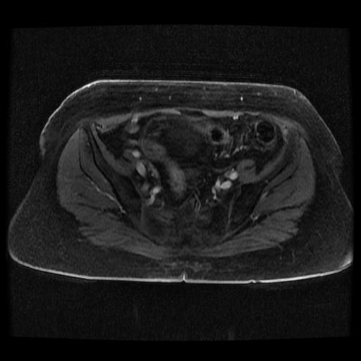
File:Cervical carcinoma (Radiopaedia 70924-81132 Axial T1 C+ fat sat 64).jpg
Jump to navigation
Jump to search
Cervical_carcinoma_(Radiopaedia_70924-81132_Axial_T1_C+_fat_sat_64).jpg (512 × 512 pixels, file size: 26 KB, MIME type: image/jpeg)
Summary:
| Description |
|
| Date | Published: 10th Sep 2019 |
| Source | https://radiopaedia.org/cases/cervical-carcinoma-2 |
| Author | Bahman Rasuli |
| Permission (Permission-reusing-text) |
http://creativecommons.org/licenses/by-nc-sa/3.0/ |
Licensing:
Attribution-NonCommercial-ShareAlike 3.0 Unported (CC BY-NC-SA 3.0)
File history
Click on a date/time to view the file as it appeared at that time.
| Date/Time | Thumbnail | Dimensions | User | Comment | |
|---|---|---|---|---|---|
| current | 11:29, 2 August 2021 |  | 512 × 512 (26 KB) | Fæ (talk | contribs) | Radiopaedia project rID:70924 (batch #7158-238 G64) |
You cannot overwrite this file.
File usage
The following page uses this file:
